Review
. 2022 Aug 31:23:29-52.
doi: 10.1146/annurev-genom-120821-100535. Epub 2022 May 24.

Extrachromosomal DNA in Cancer

Affiliations
Review

Extrachromosomal DNA in Cancer

Vineet Bafna et al. Annu Rev Genomics Hum Genet. .

Abstract

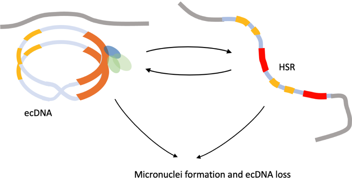
In cancer, complex genome rearrangements and other structural alterations, including the amplification of oncogenes on circular extrachromosomal DNA (ecDNA) elements, drive the formation and progression of tumors. ecDNA is a particularly challenging structural alteration. By untethering oncogenes from chromosomal constraints, it elevates oncogene copy number, drives intratumoral genetic heterogeneity, promotes rapid tumor evolution, and results in treatment resistance. The profound changes in DNA shape and nuclear architecture generated by ecDNA alter the transcriptional landscape of tumors by catalyzing new types of regulatory interactions that do not occur on chromosomes. The current suite of tools for interrogating cancer genomes is well suited for deciphering sequence but has limited ability to resolve the complex changes in DNA structure and dynamics that ecDNA generates. Here, we review the challenges of resolving ecDNA form and function and discuss the emerging tool kit for deciphering ecDNA architecture and spatial organization, including what has been learned to date about how this dramatic change in shape alters tumor development, progression, and drug resistance.

Keywords: HSRs; cancer; ecDNA; ecDNA evolution; ecDNA hubs; enhancer hijacking; homogeneously staining regions.


Conflict of interest statement

DISCLOSURE STATEMENT

VB is a co-founder, consultant, SAB member and has equity interest in Boundless Bio, inc. and Abterra, Inc. The terms of this arrangement have been reviewed and approved by the University of California, San Diego in accordance with its conflict of interest policies. P.S.M. is a co-founder of Boundless Bio, Inc. He chairs the Scientific Advisory Board, for which he is compensated.

Figures

Figure 1
Figure 1. A model of ecDNA evolution.
Bin i maintains the count Ni(t) of the number of cells with exactly i ≥ 0 ecDNA copies. At t = 1, N1(1) = 1 and all other bins are 0. An ecDNA-positive cell is s times more likely to be picked relative to an ecDNA-negative cell, where s denotes the selection coefficient. The chosen cell with i ecDNA copies replicates and divides into two daughter cells containing k and 2i − k ecDNA, chosen according to a Binomial distribution. Simulations suggest that in the absence of selection (s ≤ 1), the proportion of ecDNA-negative cells in the population increases to 1, but for s > 1, it rapidly diminishes. In the limit, the tail distribution of ecDNA counts follows a power-law with an exponential cut-off (dark black line), which is wider than the Normal distribution.
Figure 2
Figure 2. Directed assembly for ecDNA structure and detection.
(a) Short-reads from whole genome sequencing also sample ecDNA structures. (b) Paired-end sequencing and mapping identifies breakpoints and copy number changes. Colored segments represent genomic intervals of an amplicon. Gray boxes represent estimates of the copy number and multiplicities of segments (numbers). Breakpoints connecting the segments are represented by thin black lines. (c) Directed assembly methods smooth out coverage and generate an amplicon graph representing all amplified segments and their multiplicities and breakpoints. (d,e) Paths and cycles in the amplicon graph help detect ecDNA (e.g. long, high-copy cycles) and their fine structure. Large segments with high multiplicities (gray segment), missing breakpoints, or heterogeneity of ecDNA all lead to ambiguity of reconstruction. All cycles in panels d and e are consistent with the breakpoint graph.(e) Long-reads help detect breakpoints that short-reads may have missed due to low complexity sequence. Long-reads that span high multiplicity regions resolve ambiguities in reconstruction. The checked circle represents the true reconstruction.
Figure 3
Figure 3. Mechanisms of ecDNA formation.
(a) episomes formed by replication fork stalling at a bubble, DNA breakage and subsequent re-ligation leads to ecDNA formation from the one strand while repair in the second strand allows for replication to proceed without a deletion ‘scar.’ (b) Mis-segregation errors may lead to a lagging chromosome followed by micronuclei formation. Shattering in subsequent mitoses and re-ligation generate a ‘chromothriptic’ chromosome. (c) Telomere loss and sister-chromatid bridging leads to broken ends or lagging chromosomes. Repeated breakage fusion bridge cycles may lead to a rearranged chromosome with an HSR-like signature. (d) EcDNA may form by fragments breaking off and circularizing during BFB cycles or chromothripsis.
Figure 4
Figure 4. Functional characteristics of ecDNA.
(a) In contrast with chromosomes (top-panel), ecDNA (bottom-panel) is highly accessible, resulting in over-expression of genes on ecDNA. The expression level remains high even after correcting for copy number. (b) Topology Associated domains (TADs) provide the regulatory elements that control gene regulation, and shield the gene body from outside enhancers (top-panel). The circular structure of ecDNA changes the regulatory circuitry through hijacking of enhancers outside the TAD. (c) ecDNA form hubs and interact with chromosomes, resulting in enhancer activity that regulates genes in other ecDNA, and even on chromosomes.
Figure 5
Figure 5. Plasticity of ecDNA and HSR.
Aggregated ecDNA can re-combine into larger structures. Continued selection pressure that selects for amplification may lead to ecDNA formation, aggregation, and subsequent integration into a multi-copy HSR. HSRs also show plasticity and change length. Removal of selection or DNA damage leads to micronuclei formation and loss of ecDNA.

References

    1. Adelman K, Martin BJE. 2021. ecDNA party bus: Bringing the enhancer to you. Mol Cell 81(9):1866–1867 - PubMed
    1. Akagi K, Li J, Broutian TR, Padilla-Nash H, Xiao W, et al. 2014. Genome-wide analysis of HPV integration in human cancers reveals recurrent, focal genomic instability. Genome Res 24(2):185–199 - PMC - PubMed
    1. Alitalo K, Schwab M, Lin CC, Varmus HE, Bishop JM. 1983. Homogeneously staining chromosomal regions contain amplified copies of an abundantly expressed cellular oncogene (c-myc) in malignant neuroendocrine cells from a human colon carcinoma. Proc Natl Acad Sci U S A 80(6):1707–1711 - PMC - PubMed
    1. Alt FW, Kellems RE, Bertino JR, Schimke RT. 1978. Selective multiplication of dihydrofolate reductase genes in methotrexate-resistant variants of cultured murine cells. Journal of Biological Chemistry 253(5):1357–1370 - PubMed
    1. Amler LC, Schwab M. 1989. Amplified N-myc in human neuroblastoma cells is often arranged as clustered tandem repeats of differently recombined DNA. Mol Cell Biol 9(11):4903–4913 - PMC - PubMed

Publication types