

Case Reports
doi: 10.1136/bcr-2022-249828.
Sclerosing soft tissue haemangioma in an adult patient: an atypical form of a common entity causing the diagnostic dilemma
Affiliations
- PMID: 35609932
- PMCID: PMC9131102
- DOI: 10.1136/bcr-2022-249828
Item in Clipboard
Case Reports
Sclerosing soft tissue haemangioma in an adult patient: an atypical form of a common entity causing the diagnostic dilemma
BMJ Case Rep.
.
No abstract available
Keywords: Musculoskeletal and joint disorders; Orthopaedics; Radiology.
Conflict of interest statement
Competing interests: None declared.
Figures
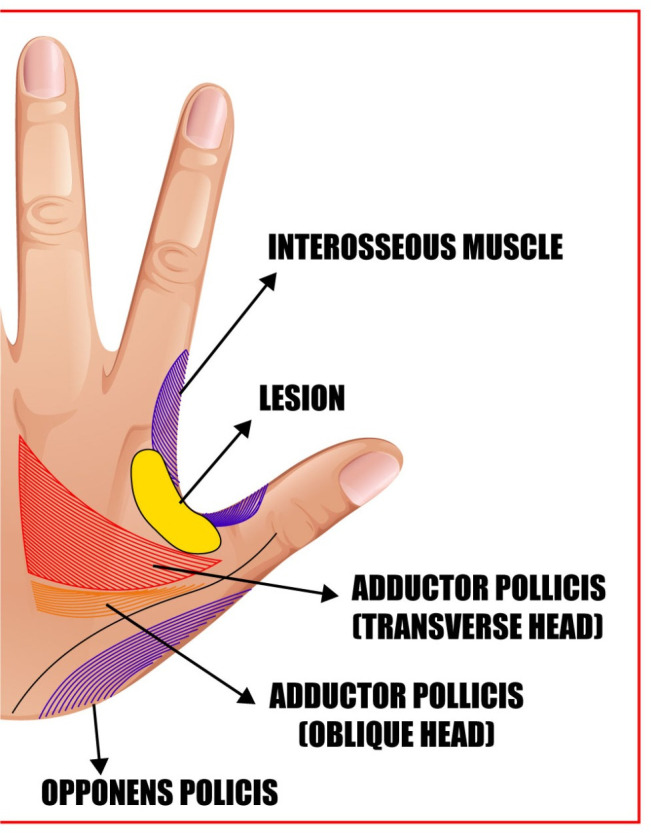
Graphic computed image of the dorsum of the left hand demonstrates location of the lesion with respect to surrounding structures (the figure has been created by the authors).
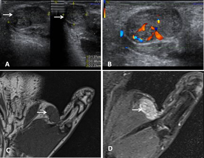
Ultrasonography still images show (A) an elongated, lobulated, well-defined, heteroechoic lesion (20×10 mm in size) in the first web space involving the subfascial and subcutaneous planes (white arrow). (B) Colour Doppler examination of the lesion demonstrates internal colour flow signals that suggest internal vascularity. (C) T1-weighted MRI shows an isointense lesion in first web space of the left hand involving subfascial and subcutaneous planes (white arrow). (D) In PDFS image, the lesion is hyperintense (white arrow) and abutting the first dorsal interosseous muscle (the figures have been created by the authors). PDFS, proton density fat suppressed; USG, ultrasonography.
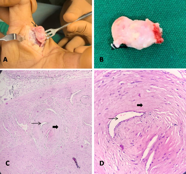
(A, B) Intraoperative and gross specimen images showing an almond-sized lesion excised from first web space of the hand. (C, D) Histopathology images with 10× and 40× magnifications show multiple dense whirls of hyalinised stroma obliterating the endothelium-lined proliferated vascular channels, suggestive of sclerosing variety of haemangioma (the figures have been created by the authors).
Similar articles
-
Progressively growing paediatric knee swelling: synovial haemangioma.BMJ Case Rep. 2021 Sep 22;14(9):e242694. doi: 10.1136/bcr-2021-242694. BMJ Case Rep. 2021. PMID: 34551911 Free PMC article. No abstract available.
-
Cytological imprint in frozen section diagnosis of sclerosing haemangioma of the lung: a case report.Mater Med Pol. 1994 Apr-Jun;26(2):73-4. Mater Med Pol. 1994. PMID: 7745988
-
Unusual sclerosing haemangiomas and sclerosing haemangioma-like lesions, and the value of TTF-1 in making the diagnosis.Histopathology. 2002 Nov;41(5):404-13. doi: 10.1046/j.1365-2559.2002.01522.x. Histopathology. 2002. PMID: 12405908
-
Sclerosing haemangioma of the lung.Histol Histopathol. 1992 Apr;7(2):209-12. Histol Histopathol. 1992. PMID: 1515703 Review.
-
Sclerosing hemangioma of the lung.Arch Pathol Lab Med. 2009 May;133(5):820-5. doi: 10.5858/133.5.820. Arch Pathol Lab Med. 2009. PMID: 19415961 Review.
References
Publication types
MeSH terms
LinkOut - more resources
Full Text Sources
Medical