. 2022 Jun 6;9(3):ENEURO.0373-21.2022.
doi: 10.1523/ENEURO.0373-21.2022. Print 2022 May-Jun.

Selectively Imaging Cranial Sensory Ganglion Neurons Using AAV-PHP.S

Affiliations

Selectively Imaging Cranial Sensory Ganglion Neurons Using AAV-PHP.S

Andoni I Asencor et al. eNeuro. .

Abstract

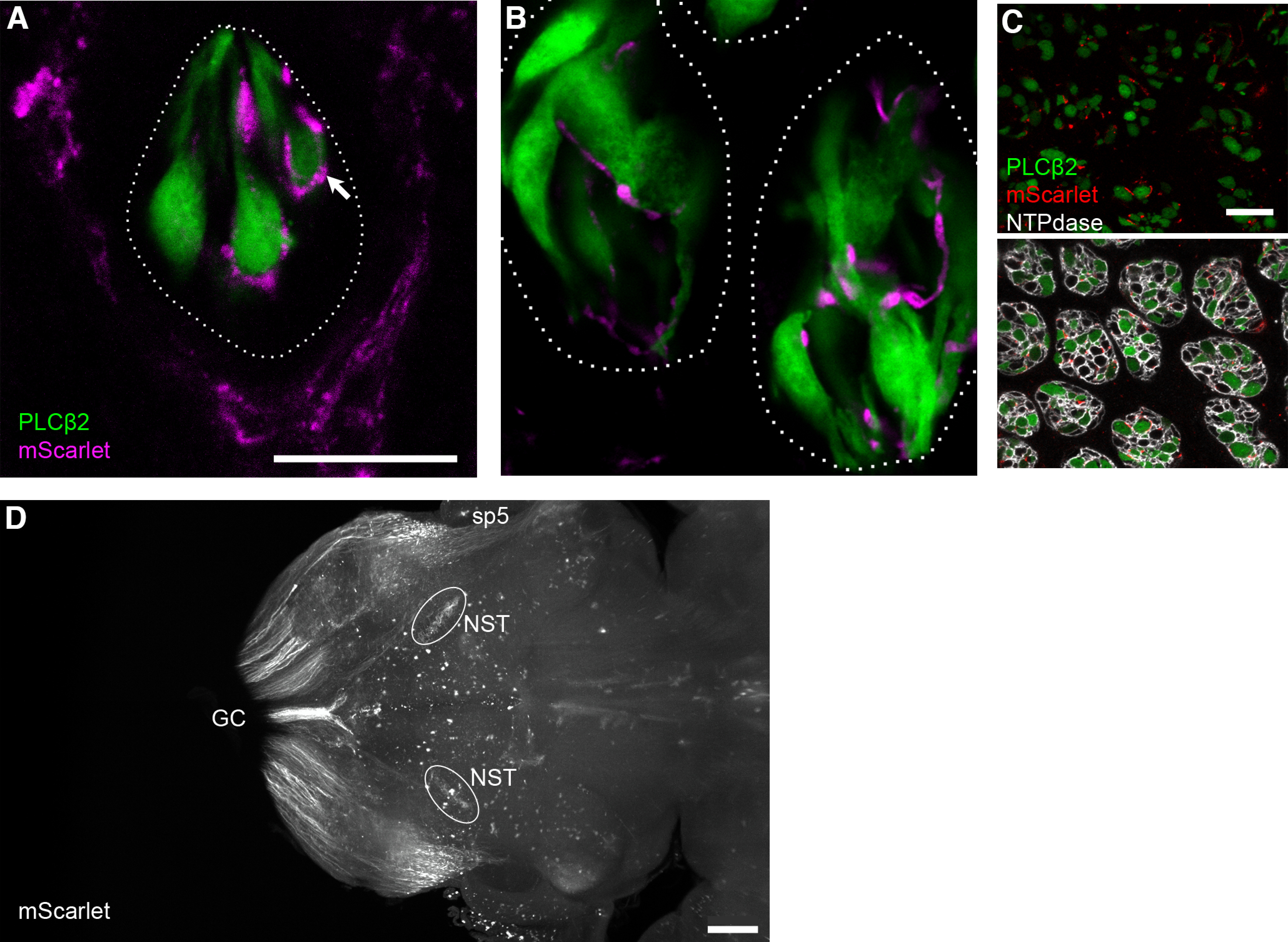
Because of their ease of use, adeno-associated viruses (AAVs) are indispensable tools for much of neuroscience. Yet AAVs have been used relatively little to study the identities and connectivity of peripheral sensory neurons, principally because methods to selectively target peripheral neurons have been limited. The introduction of the AAV-PHP.S capsid with enhanced tropism for peripheral neurons (Chan et al., 2017) offered a solution, which we further elaborate here. Using AAV-PHP.S with GFP or mScarlet fluorescent proteins, we show that the mouse sensory ganglia for cranial nerves V, VII, IX, and X are targeted. Pseudounipolar neurons of both somatic and visceral origin, but not satellite glia, express the reporters. One week after virus injection, ≈66% of geniculate ganglion neurons were transduced. Fluorescent reporters were transported along the central and peripheral axons of these sensory neurons, permitting visualization of terminals at high resolution, and in intact, cleared brain using light sheet microscopy. Further, using a Cre-dependent reporter, we demonstrate by anatomic and functional criteria, that expression is in a cell type-selective manner. Finally, we integrate earlier neuroanatomical and molecular data with in vivo Ca2+ imaging to demonstrate the sensory characteristics of geniculate ganglion auricular neurons, which were previously undocumented. Our analyses suggest that the AAV-PHP.S serotype will be a powerful tool for anatomically and functionally mapping the receptive fields and circuits of the expanding numbers of molecular subtypes of many somatosensory and viscerosensory neurons that continue to be defined via single-cell RNA sequencing.

Keywords: AAV-PHP.S; auricular neurons; calcium imaging; labeling afferent fibers; pseudounipolar sensory neurons; somatosensory.


Figures

Figure 1.
Figure 1.
AAV-PHP.S transduces neurons in multiple sensory ganglia. AAV-PHP.S::CAG-GFP, injected into the retroorbital sinus of three mice, resulted in expression of GFP in (A) dorsal root, (B) nodose-petrosal complex, (C) geniculate, and (D) trigeminal ganglia, viewed for GFP intrinsic fluorescence in cryosections. Ganglia were dissected 7 d postinjection. E, Cryosections of trigeminal ganglion (as in D) were immunostained for NeuN (magenta) and GFAP (orange) to identify neurons and satellite glia, respectively. Only neurons are seen to express GFP. F, Cryosections of geniculate ganglion (as in C) were immunostained for Phox2b (orange) and Drg11 (magenta) to identify visceral and somatic neuronal nuclei, respectively. Several neurons of each class (arrowheads, somatic; arrows, visceral) are seen to express GFP (green). All images are single confocal plane. Scale bars: 50 μm. A small number of GFP+ cells were also observed in cochlea (Extended Data Fig. 1-1).
Figure 2.
Figure 2.
Time course of GFP expression. A–E, Confocal images of cryosections of geniculate ganglia from mice injected with AAV-PHP.S::CAG-GFP, and analyzed at 2, 4, 7, 14, and 21 d postinjection. Intrinsic fluorescence of GFP was captured in parallel for all images; brightness was increased only in A to demonstrate very low-level expression at the earliest stage. F, Transduction efficiency was quantified in confocal images of cryosections immunostained for NeuN. All neurons that displayed fluorescence visibly above background on the facial nerve (fn) track were scored as positive (solid arrowhead in D). An example of a GFP-negative is indicated (open arrowhead in D). Data are presented as the percentage of neurons that were GFP-labeled. Each symbol represents data from 400–600 neurons from a separate mouse (with 2, 5, 3, 3, 3 mice at successive time points). Solid line is the average for all mice at each time point. Scale bar: 50 μm.
Figure 3.
Figure 3.
Only PNS neurons are targeted; CNS neurons remain unlabeled. Mice were injected with AAV-PHP.S::CAG-GFP and tissues were harvested 14 d later. A, Cryosection of hindbrain, immunostained for NeuN (orange) shows that AAV-delivered GFP is absent from central neurons and limited to areas that receive afferent inputs from peripheral sensory neurons: the spinal trigeminal tract (sp5), spinal trigeminal nucleus interpolar (Sp5l) and rNST with its gustatory inputs. B, Section of mid-thoracic spinal cord, imaged for NeuN immunoreactivity (magenta) and intrinsic fluorescence of AAV-delivered GFP, which is limited to sensory inputs from the DRG. C, Higher magnification of boxed region of rNST, additionally stained for P2X3 (magenta) to label gustatory afferents. Many of the finest GFP+ fibers are gustatory, with boutons that suggest synapses on rNST neurons (arrowheads). D, High magnification of a region of ventral horn indicated by a box in B, where GFP+ afferent sensory fibers present boutons resembling synapses, juxtaposed against primary motor neurons (identified by their large size). Scale bars: 500 μm (A, B) and 50 μm (C, D).
Figure 4.
Figure 4.
Peripheral terminals of sensory ganglion neurons are GFP-labeled. A–F, Cryosections of fungiform (A, C, E) and circumvallate (B, D, F) taste buds from nine mice, injected with AAV-PHP.S;;CAG-GFP, were immunostained 7, 14, or 21 d postinjection to detect accumulation of axonally transported GFP to the peripheral terminals of sensory ganglion neurons. Dotted lines outline each taste buds (38–62 analyzed per mouse). Gustatory fibers are immunoreactive for P2X3 (magenta), allowing discrimination from P2X3-negative trigeminal fibers outside the taste buds. The number of GFP+ fibers visible and their fluorescence intensity appears to increase from 7 to 21 d. All images were captured in parallel at the same settings to make fluorescence intensities comparable. G, H, Quantification of incidence of GFP+ fibers within perimeter of taste buds at each time point. Each green symbol represents the fraction of 15–29 fungiform taste buds from one mouse (G) or 23–33 circumvallate taste buds from one mouse (H). Data from nine mice are included in each graph. In G, the secondary y-axis depicts total fluorescence intensity of GFP+ fibers within each fungiform taste bud across the time course from 7 to 21 d. Each gray data point is a separate taste bud; a total of 20 fungiform taste buds from five mice were sampled for gray symbols. Scale bar: 20 μm.
Figure 5.
Figure 5.
mScarlet reporter for visualizing individual fibers and fiber tracts. A–C, Fungiform and circumvallate taste buds from a Plcb2-GFP mouse, injected with AAV-PHP.S::CAG-mScarlet-I, examined 21 d postinjection. In a single confocal plane of a fungiform papilla (A), two types of mScarlet+ nerves (magenta) are visible: trigeminal fibers that form a corona around the taste bud, and gustatory fibers that enter the taste bud. Terminal boutons of gustatory nerves are seen associated with individual Type II (GFP-expressing, green) chemoreceptor cells (arrow). B, In a single confocal plane, two circumvallate taste buds similarly show gustatory afferents associating with GFP+ chemosensory cells. C, Circumvallate taste buds from the same mouse as in A, B, viewed in cross-section and at low magnification, nearly every taste bud includes multiple afferent fibers that are mScarlet-labeled (red). Immunoreactivity for NTPDase2 outlines each taste bud cell. D, Light-sheet microscope imaging of a cleared brain of a mouse injected with AAV-PHP.S;;CAG-mScarlet-I. Brain was viewed in a horizontal orientation. This image is a Z-projection through a 100-μm thickness that includes the rNST. The sensory afferent fibers of AAV-transduced neurons detected here include the gracile and cuneate sensory tracts (GC) entering the medulla caudally, the spinal trigeminal tract (sp5) and the terminals of gustatory afferents in the rNST. Scale bar: 20 μm (for taste buds) and 500 μm (for brain). A 3D reconstruction of the mScarlet-labeled hindbrain is in Movie 1, and permits visualization of the entry of multiple sensory afferents.
Figure 6.
Figure 6.
Ca2+ imaging of gustatory neurons with Cre-dependent GCaMP in a Penk-Cre mouse. Geniculate ganglion neurons were imaged under anesthesia 14 d after injection with AAV-PHP.S::flex-GCaMP6s. Traces are the responses (ΔF/F0) of four representative GCaMP+ neurons to oral perfusion of three acidic taste stimuli (gray bars): citric acid (ca), HCl (ha), acetic acid (aa), followed by mechano-stimulation of the whiskers (w) or pinna (as in Fig. 7) with a puff of compressed air (a), stroking with a bristle brush (b), and a flick (f). None of the mechanical stimuli elicited a response. Scale bar: 10 s, 1.0 ΔF/F0.
Figure 7.
Figure 7.
Cre-dependent expression in peripheral sensory neurons of a Mafb-mCherry-Cre mouse. The geniculate ganglion was examined 14 d after injecting AAV-PHP.S::flex-GCaMP6s. Cryosections were immunostained with anti-GFP (to detect GCaMP), anti-mCherry, and anti-Phox2b (to detect gustatory neurons). GCaMP6 (green) is detected only in neurons that express Cre and mCherry (red, filled arrowhead). The overwhelming majority of gustatory neurons in the ganglion lack mCherry and Cre, have Phox2b+ nuclei and did not express GCaMP (open arrowhead). Scale bar: 50 μm. The lack of GCaMP expression in central neurons that express Cre is shown in Extended Data Figure 7-1.
Figure 8.
Figure 8.
Ca2+ imaging of auricular neurons with Cre-dependent GCaMP in Mafb-mCherry-Cre mouse. A, Geniculate ganglion of anesthetized mouse, 14 d after injection with AAV-PHP.S::flex-GCaMP6s, viewed for mCherry (red) and GCaMP (green) during recording. This region of the ganglion includes a high density of auricular (mCherry+) neurons, many of which express GCaMP. B, Responses (ΔF/F0) of 6 GCaMP+ auricular neurons from a different ganglion, similar to A, to five types of mechano-stimulation of the rigid, cartilaginous portion of the pinna. Stimuli (gray bars) included from left to right, a puff of compressed air (a), stroking with a bristle brush (b), gentle touch with flat spatula (t), deep pressure with same spatula (p), and deflection of pinna with same spatula (d). Example traces of two neurons that responded in each of three different patterns are shown. C, Responses of two GCaMP+ auricular neurons, stimulated first with mechanical stimuli as in B, then by flicking the pinna (f) or whiskers (w), and finally with oral perfusion of five stereotypical taste stimuli in the mouth. Zero of 49 neurons stimulated in this manner (across 4 mice) responded to whisker stimulation or oral tastants: sucrose (s), NaCl (n), citric acid (ca), quinine/cycloheximide (q) and MSG/IMP (m). Scale bars for B, C: 10 s, 1.0 ΔF/F0.

References

    1. Aschauer DF, Kreuz S, Rumpel S (2013) Analysis of transduction efficiency, tropism and axonal transport of AAV serotypes 1, 2, 5, 6, 8 and 9 in the mouse brain. PLoS One 8:e76310. 10.1371/journal.pone.0076310 - DOI - PMC - PubMed
    1. Bartel DL, Sullivan SL, Lavoie EG, Sévigny J, Finger TE (2006) Nucleoside triphosphate diphosphohydrolase-2 is the ecto-ATPase of type I cells in taste buds. J Comp Neurol 497:1–12. 10.1002/cne.20954 - DOI - PMC - PubMed
    1. Bas E, Anwar MR, Goncalves S, Dinh CT, Bracho OR, Chiossone JA, Van De Water TR (2019) Laminin-coated electrodes improve cochlear implant function and post-insertion neuronal survival. Neuroscience 410:97–107. 10.1016/j.neuroscience.2019.04.048 - DOI - PubMed
    1. Bindels DS, Haarbosch L, van Weeren L, Postma M, Wiese KE, Mastop M, Aumonier S, Gotthard G, Royant A, Hink MA, Gadella TW Jr (2017) mScarlet: a bright monomeric red fluorescent protein for cellular imaging. Nat Methods 14:53–56. 10.1038/nmeth.4074 - DOI - PubMed
    1. Bloom DC, Watson ZL, Neumann DM (2019) Peripheral AAV injection for retrograde transduction of dorsal root and trigeminal ganglia. Methods Mol Biol 1950:237–247. - PubMed

Publication types

LinkOut - more resources