. 2022 May 24;13(5):493.
doi: 10.1038/s41419-022-04957-9.

Disruption of β-catenin-mediated negative feedback reinforces cAMP-induced neuronal differentiation in glioma stem cells

Affiliations

Disruption of β-catenin-mediated negative feedback reinforces cAMP-induced neuronal differentiation in glioma stem cells

Zhijie Chen et al. Cell Death Dis. .

Abstract

Accumulating evidence supports the existence of glioma stem cells (GSCs) and their critical role in the resistance to conventional treatments for glioblastoma multiforme (GBM). Differentiation therapy represents a promising alternative strategy against GBM by forcing GSCs to exit the cell cycle and reach terminal differentiation. In this study, we demonstrated that cAMP triggered neuronal differentiation and compromised the self-renewal capacity in GSCs. In addition, cAMP induced negative feedback to antagonize the differentiation process by activating β-catenin pathway. Suppression of β-catenin signaling synergized with cAMP activators to eliminate GSCs in vitro and extended the survival of animals in vivo. The cAMP/PKA pathway stabilized β-catenin through direct phosphorylation of the molecule and inhibition of GSK-3β. The activated β-catenin translocated into the nucleus and promoted the transcription of APELA and CARD16, which were found to be responsible for the repression of cAMP-induced differentiation in GSCs. Overall, our findings identified a negative feedback mechanism for cAMP-induced differentiation in GSCs and provided potential targets for the reinforcement of differentiation therapy for GBM.


Conflict of interest statement

The authors declare no competing interests.

Figures
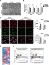
Fig. 1
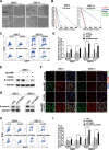
Fig. 1. cAMP induces neuronal differentiation in GSCs.
A Phase-contrast image of GSC1 and GSC11 cells before and after treatment with dbcAMP and forskolin for 72 h (scale bar, 100 μm). B Quantification of MAP2 and TUJ1 in GSCs treated with dbcAMP and forskolin by flow cytometry. C Immunofluorescence staining of MAP2 (red) and TUJ1 (green) in GSCs before and after incubation with dbcAMP (left) and quantification of fluorescence intensity (right) (scale bar, 50 μm). D Heat map of differentially expressed genes between GSCs treated with and without dbcAMP. E GSEA analysis using the mRNA sequencing data. (Data are shown as the mean ± SD. N.S., not significant; *, p < 0.05; **, p < 0.01; ***, p < 0.001; ****, p < 0.0001).
Fig. 2
Fig. 2. Activation of cAMP compromises self-renewal and represses proliferation in GSCs.
A Phase-contrast images of neurospheres formed by GSC1 and GSC11 cells after treatment with cAMP activators for 72 h. B Limiting dilution assays of GSC1 and GSC11 cells treated with dbcAMP and forskolin. C Western blot of CD133 and SOX2 protein in GSCs exposed to dbcAMP and forskolin (left) and quantification of protein bands (right). D GSEA analysis using the mRNA sequencing data. E Flow cytometry and EdU incorporation DNA histograms of GSCs before and after the treatment with dbcAMP and forskolin. (scale bar, 100 μm; data are shown as the mean ± SD. N.S., not significant; *, p < 0.05; **, p < 0.01; ***, p < 0.001; ****, p < 0.0001).
Fig. 3
Fig. 3. cAMP activates β-catenin signaling by inhibiting destabilization of β-catenin through PKA and GSK-3β.
A Heat map of DEGs between GSCs treated with and without dbcAMP. B GSEA analysis using the mRNA sequencing data. C Western blot of β-catenin protein in GSCs incubated with dbcAMP and forskolin. D Immunofluorescence staining of β-catenin (green) in GSCs treated with dbcAMP at 0 (control), 24, 48, and 72 h. E Western blot of total and phosphorylated β-catenin in GSCs subjected to different treatments. F Protein level of phosphorylated GSK-3β at Ser9 and GSK-3β assessed by Western blot in GSCs treated with dbcAMP at 0, 24, 48, and 72 h. (Scale bar, 25 μm).
Fig. 4
Fig. 4. Suppression of β-catenin promotes cAMP-induced differentiation in GSCs.
A Phase-contrast image of neurospheres formed by GSC1 and GSC11 cells after various treatments for 72 h (scale bar, 100 μm). B Limiting dilution assays of GSC1 and GSC11cells treated with dbcAMP, FH535, and the combination of dbcAMP and FH535. C Proliferation was evaluated by EdU incorporation analysis in GSCs treated with dbcAMP, FH535, and the combination of the two agents. D Western blot of β-catenin in GSC1 treated with dbcAMP and FH535. E Quantification by flow cytometry of MAP2 and TUJ1 in GSCs incubated with dbcAMP and FH535. F Immunofluorescence staining of MAP2 (red) and TUJ1 (green) in GSCs with different treatments (scale bar, 50 μm). G Western blot of β-catenin in GSCs treated with siRNA specific for β-catenin (siCTNNB1). H Proliferation evaluated by EdU incorporation analysis of GSCs with β-catenin knock-down. I Quantification of MAP2 and TUJ1 in GSCs with β-catenin knock-down by using flow cytometry. (Data are shown as the mean ± SD. N.S., not significant; *, p < 0.05; **, p < 0.01; ***, p < 0.001).
Fig. 5
Fig. 5. β-catenin inhibitor synergizes with cAMP agonists in GSC xenograft models.
A Tumor growth curve of subcutaneous mouse model treated with cAMP activators and FH535. B Representative photos of subcutaneous tumor specimens in different groups. C Changes of the body weights after treatment in various groups. D Representative images of mouse brain specimens from orthotopic models treated with vehicle, dbcAMP+Luteolin (dL), FH535, dL+FH535. E Kaplan–Meier curve showing survival of mice bearing GSCs treated with cAMP activators, FH535 and the combination. F H&E staining of tumors derived from GSCs subjected to different treatments. Tumor tissues were stained with antibodies against Ki-67 and MAP2. (Scale bar, 25 μm; Data are shown as the mean ± SD. N.S., not significant; *, p < 0.05; **, p < 0.01; ***, p < 0.001).
Fig. 6
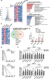
Fig. 6. Identification of β-catenin downstream genes that contribute to the negative regulation of cAMP-induced differentiation in GSCs.
A Volcano plot and heat map show the distribution of DEGs in dbcAMP-treated GSCs before and after the knock-down of β-catenin. B Function and pathway enrichment of DEGs in dbcAMP-treated GSCs before and after the knock-down of β-catenin. C Heat map and Venn diagram demonstrating the overlap of top downregulated DEGs in the two gene sets. D Validation of the expression of four DEGs by using qRT-PCR. E Limiting dilution assays of GSC1 and GSC11 cells after treatment with specific siRNA targeting APELA, CARD16 and MSLN. F Quantification of MAP2 and TUJ1 in GSCs treated with the combination of dbcAMP and knock-down of CTNNB1, APELA, CARD16 or MSLN, by using flow cytometry. (db, dbcAMP; Data are shown as the mean ± SD. N.S., not significant; *, p < 0.05; **, p < 0.01; ***, p < 0.001).
Fig. 7
Fig. 7
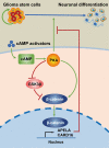
The schematic diagram shows the proposed mechanism that cAMP activates β-catenin through PKA and GSK3β, and afterwards promotes transcription of the downstream genes APELA and CARD16, which eventually prevent neuronal differentiation in GSCs.

References

    1. Wen PY, Weller M, Lee EQ, Alexander BM, Barnholtz-Sloan JS, Barthel FP, et al. Glioblastoma in adults: A Society for Neuro-Oncology (SNO) and European Society of Neuro-Oncology (EANO) consensus review on current management and future directions. Neuro Oncol. 2020;22:1073–113. doi: 10.1093/neuonc/noaa106. - DOI - PMC - PubMed
    1. Neftel C, Laffy J, Filbin MG, Hara T, Shore ME, Rahme GJ, et al. An integrative model of cellular states, plasticity, and genetics for glioblastoma. Cell. 2019;178:835–49 e21. doi: 10.1016/j.cell.2019.06.024. - DOI - PMC - PubMed
    1. Lathia JD, Mack SC, Mulkearns-Hubert EE, Valentim CL, Rich JN. Cancer stem cells in glioblastoma. Genes Dev. 2015;29:1203–17. doi: 10.1101/gad.261982.115. - DOI - PMC - PubMed
    1. Singh SK, Clarke ID, Terasaki M, Bonn VE, Hawkins C, Squire J, et al. Identification of a cancer stem cell in human brain tumors. Cancer Res. 2003;63:5821–8. - PubMed
    1. Galli R, Binda E, Orfanelli U, Cipelletti B, Gritti A, De Vitis S, et al. Isolation and characterization of tumorigenic, stem-like neural precursors from human glioblastoma. Cancer Res. 2004;64:7011–21. doi: 10.1158/0008-5472.CAN-04-1364. - DOI - PubMed

Publication types

Substances