. 2022 May 24;19(1):119.
doi: 10.1186/s12974-022-02476-0.

Network alterations underlying anxiety symptoms in early multiple sclerosis

Affiliations

Network alterations underlying anxiety symptoms in early multiple sclerosis

Erik Ellwardt et al. J Neuroinflammation. .

Abstract

Background: Anxiety, often seen as comorbidity in multiple sclerosis (MS), is a frequent neuropsychiatric symptom and essentially affects the overall disease burden. Here, we aimed to decipher anxiety-related networks functionally connected to atrophied areas in patients suffering from MS.

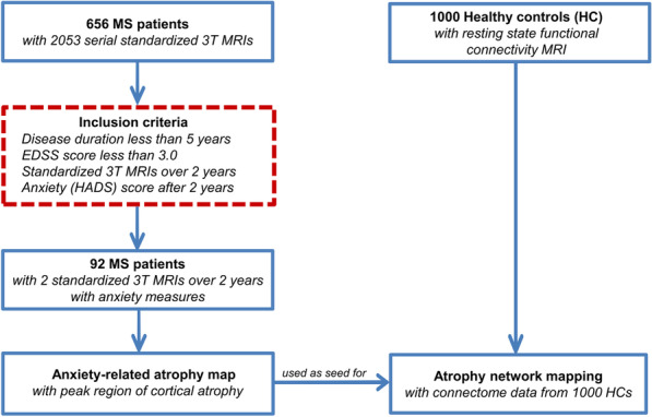
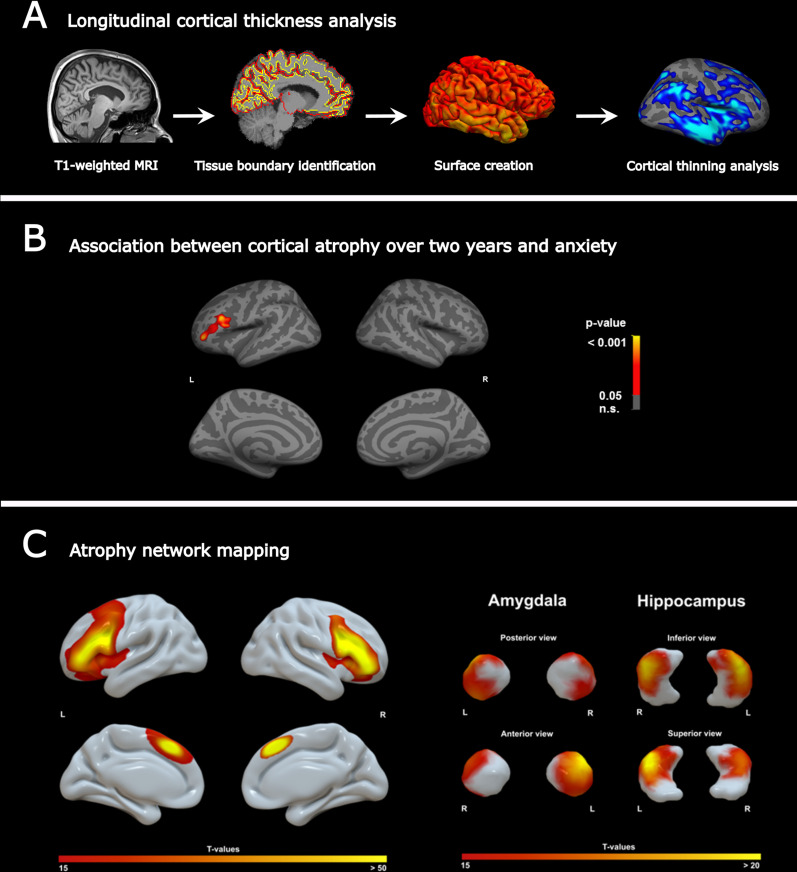
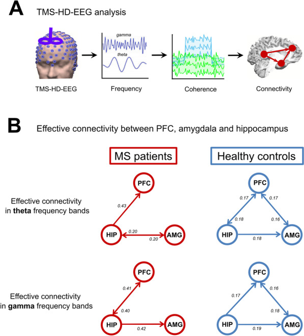
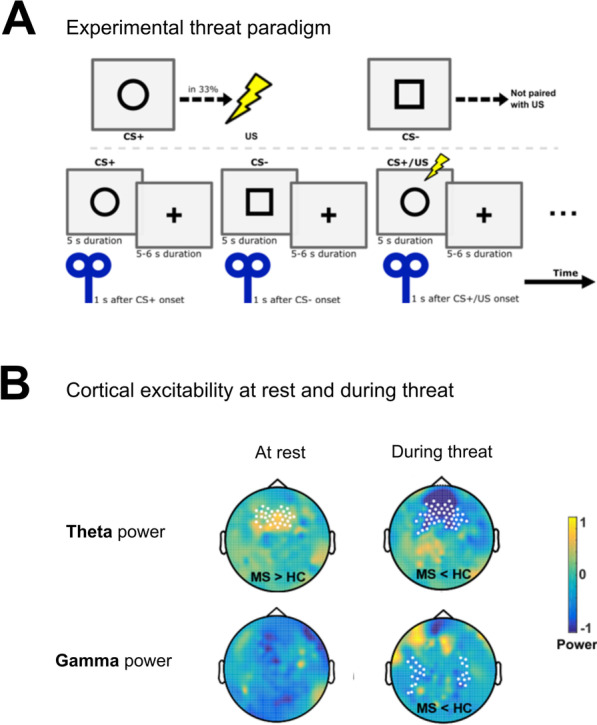
Methods: Using 3-T MRI, anxiety-related atrophy maps were generated by correlating longitudinal cortical thinning with the severity of anxiety symptoms in MS patients. To determine brain regions functionally connected to these maps, we applied a technique termed "atrophy network mapping". Thereby, the anxiety-related atrophy maps were projected onto a large normative connectome (n = 1000) performing seed-based functional connectivity. Finally, an instructed threat paradigm was conducted with regard to neural excitability and effective connectivity, using transcranial magnetic stimulation combined with high-density electroencephalography.

Results: Thinning of the left dorsal prefrontal cortex was the only region that was associated with higher anxiety levels. Atrophy network mapping identified functional involvement of bilateral prefrontal cortex as well as amygdala and hippocampus. Structural equation modeling confirmed that the volumes of these brain regions were significant determinants that influence anxiety symptoms in MS. We additionally identified reduced information flow between the prefrontal cortex and the amygdala at rest, and pathologically increased excitability in the prefrontal cortex in MS patients as compared to controls.

Conclusion: Anxiety-related prefrontal cortical atrophy in MS leads to a specific network alteration involving structures that resemble known neurobiological anxiety circuits. These findings elucidate the emergence of anxiety as part of the disease pathology and might ultimately enable targeted treatment approaches modulating brain networks in MS.

Keywords: Anxiety; Atrophy; Excitability; Functional connectivity; Multiple sclerosis.


Conflict of interest statement

Stefan Bittner has recently received consultation funds from Biogen Idec, Merck Serono, Novartis, Sanofi-Genzyme and Roche. Frauke Zipp has recently received research grants and/or consultation funds from the DFG, BMBF, PMSA, Genzyme, Janssen, Merck Serono, Roche, Novartis, Celgene, Sanofi-Aventis. The other authors report no relevant competing interests.

Figures

Fig. 1
Fig. 1
Study design and work flow. After fulfilling the inclusion criteria (red dashed line box), 92 early multiple sclerosis patients were included for the cortical thickness correlation analysis with HADS-A scores (anxiety symptoms) to generate an anxiety-related atrophy map. Next, atrophy network mapping was applied to project this atrophy map onto a normative functional brain network (n = 1000). As a result, we were able to determine brain regions functionally connected to the atrophied location
Fig. 2
Fig. 2
A Longitudinal cortical thickness analysis. Workflow for longitudinal MRI morphometric analysis. B Anxiety-related atrophy maps across multiple sclerosis patients. Atrophy of the left PFC (peak region: rostral middle frontal lobe) was associated with anxiety scores measured by the HADS-A scale. C Atrophy network mapping. Generated atrophy maps are used in a seed-based functional connectivity analysis in a large dataset of healthy controls to find functionally connected regions. The atrophy network mapping approach identified the PFC, amygdala and hippocampus as brain regions functionally connected to the previously detected atrophied location
Fig. 3
Fig. 3
Impaired connectivity between anxiety-related regions in multiple sclerosis patients. A In a second patient cohort (n = 18) TMS–HD-EEG was performed at rest and effective connectivity between the PFC, amygdala and hippocampus was measured and compared to healthy controls (n = 18). B Theta and gamma frequency bands revealed no connectivity between PFC and amygdala in patients versus controls
Fig. 4
Fig. 4
Altered cortical excitability in multiple sclerosis in response to threat. TMS–HD-EEG was performed in a second patient cohort (n = 18) and compared to healthy controls (n = 18). A Threat paradigm with a 33% chance of receiving a shock after appearance of a circle displayed on a monitor. A TMS pulse was applied 1 s after cue onset. HD-EEG (256 electrodes) was performed simultaneously. B Multiple sclerosis versus control showed an increased theta power at rest in prefrontal regions following TMS stimulation of the dorsal PFC; under threat, multiple sclerosis patients displayed a reduced theta and gamma power

References

    1. Calabrese P, Penner IK. Cognitive dysfunctions in multiple sclerosis—a “multiple disconnection syndrome”? J Neurol. 2007;254(Suppl 2):18–21. - PubMed
    1. Murphy R, O'Donoghue S, Counihan T, McDonald C, Calabresi PA, Ahmed MA, et al. Neuropsychiatric syndromes of multiple sclerosis. J Neurol Neurosurg Psychiatry. 2017;88(8):697–708. doi: 10.1136/jnnp-2016-315367. - DOI - PubMed
    1. Caceres F, Vanotti S, Benedict RH, Group RW Cognitive and neuropsychiatric disorders among multiple sclerosis patients from Latin America: results of the RELACCEM study. Mult Scler Relat Disord. 2014;3(3):335–340. doi: 10.1016/j.msard.2013.10.007. - DOI - PubMed
    1. Pape K, Tamouza R, Leboyer M, Zipp F. Immunoneuropsychiatry—novel perspectives on brain disorders. Nat Rev Neurol. 2019;15(6):317–328. doi: 10.1038/s41582-019-0174-4. - DOI - PubMed
    1. Korostil M, Feinstein A. Anxiety disorders and their clinical correlates in multiple sclerosis patients. Mult Scler. 2007;13(1):67–72. doi: 10.1177/1352458506071161. - DOI - PubMed