. 2022 Jul 8;131(2):e34-e50.
doi: 10.1161/CIRCRESAHA.121.319976. Epub 2022 May 25.

Targeting Adiponectin Receptor 1 Phosphorylation Against Ischemic Heart Failure

Affiliations

Targeting Adiponectin Receptor 1 Phosphorylation Against Ischemic Heart Failure

Di Zhu et al. Circ Res. .

Abstract

Background: Despite significantly reduced acute myocardial infarction (MI) mortality in recent years, ischemic heart failure continues to escalate. Therapeutic interventions effectively reversing pathological remodeling are an urgent unmet medical need. We recently demonstrated that AdipoR1 (APN [adiponectin] receptor 1) phosphorylation by GRK2 (G-protein-coupled receptor kinase 2) contributes to maladaptive remodeling in the ischemic heart. The current study clarified the underlying mechanisms leading to AdipoR1 phosphorylative desensitization and investigated whether blocking AdipoR1 phosphorylation may restore its protective signaling, reversing post-MI remodeling.

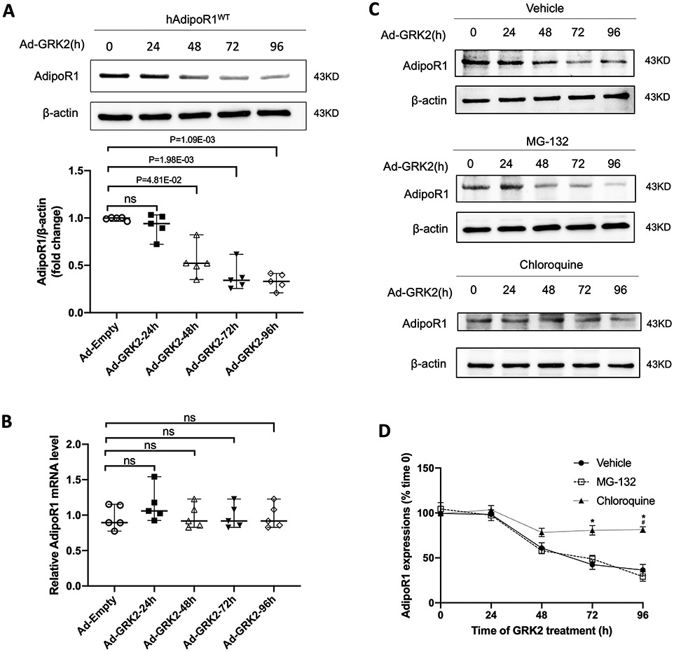
Methods: Specific sites and underlying molecular mechanisms responsible for AdipoR1 phosphorylative desensitization were investigated in vitro (neonatal and adult cardiomyocytes). The effects of AdipoR1 phosphorylation inhibition upon APN post-MI remodeling and heart failure progression were investigated in vivo.

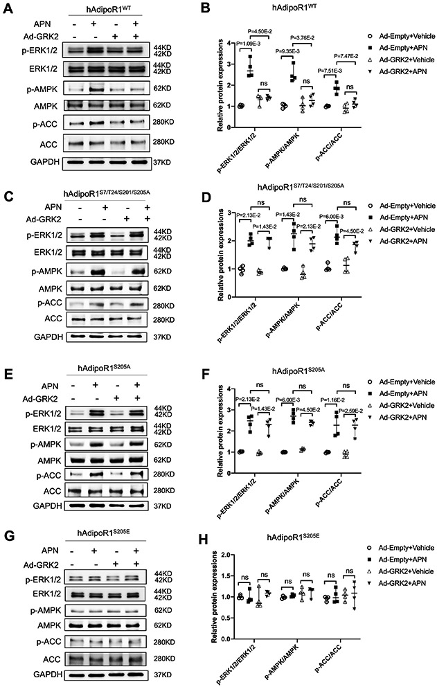
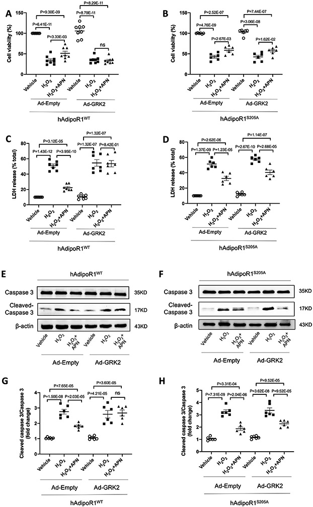
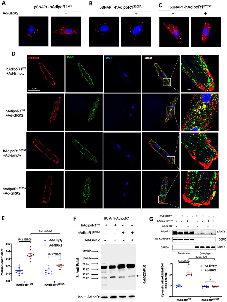
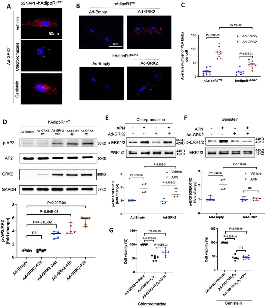
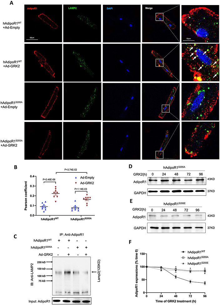
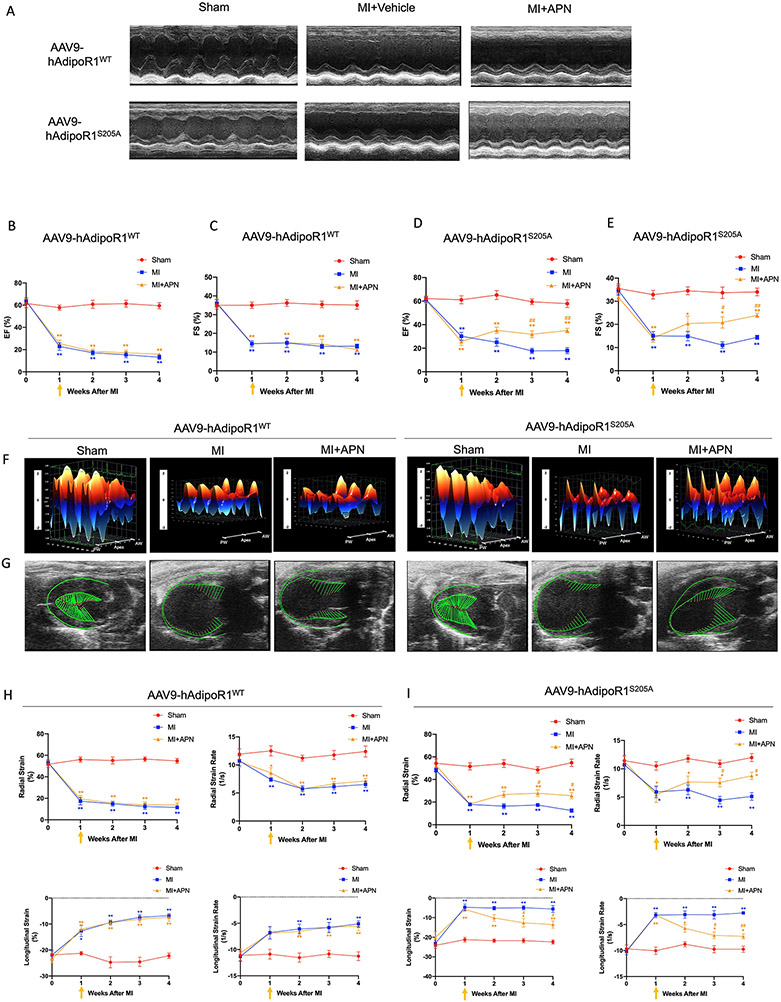
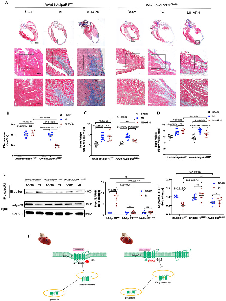
Results: Among 4 previously identified sites sensitive to GRK2 phosphorylation, alanine substitution of Ser205 (AdipoR1S205A), but not other 3 sites, rescued GRK2-suppressed AdipoR1 functions, restoring APN-induced cell salvage kinase activation and reducing oxidative cell death. The molecular investigation followed by functional determination demonstrated that AdipoR1 phosphorylation promoted clathrin-dependent (not caveolae) endocytosis and lysosomal-mediated (not proteasome) degradation, reducing AdipoR1 protein level and suppressing AdipoR1-mediated cytoprotective action. GRK2-induced AdipoR1 endocytosis and degradation were blocked by AdipoR1S205A overexpression. Moreover, AdipoR1S205E (pseudophosphorylation) phenocopied GRK2 effects, promoted AdipoR1 endocytosis and degradation, and inhibited AdipoR1 biological function. Most importantly, AdipoR1 function was preserved during heart failure development in AdipoR1-KO (AdipoR1 knockout) mice reexpressing hAdipoR1S205A. APN administration in the failing heart reversed post-MI remodeling and improved cardiac function. However, reexpressing hAdipoR1WT in AdipoR1-KO mice failed to restore APN cardioprotection.

Conclusions: Ser205 is responsible for AdipoR1 phosphorylative desensitization in the failing heart. Blockade of AdipoR1 phosphorylation followed by pharmacological APN administration is a novel therapy effective in reversing post-MI remodeling and mitigating heart failure progression.

Keywords: adipokines; animals; cell death; endocytosis; heart failure.


Figures

Figure 1.
Figure 1.
Ser205 phosphorylation is responsible for AdipoR1 desensitization and blocking AdipoR1-mediated transmembrane signaling. NMVMs isolated from adipoR1 knock out mice were transfected with 3xFlag-hAdipoR1WT, hAdipoR1S7/T24/S201/S205A, hAdipoR1S205A or hAdipoR1S205E. 24 hours after transfection, cells were infected with either an Ad-empty or Ad-GRK2 vector. 48 hours later, cells were treated with APN (2 μg/mL) for 30 minutes. A/C/E/G: Effect of APN on cell salvage kinase activation in AdipoR1-KO/hAdipoR1WT, AdipoR1-KO/hAdipoR1S7/T24/S201/S205A, AdipoR1-KO/hAdipoR1S205A or AdipoR1-KO/hAdipoR1S205E cardiomyocytes. B/D/F/H: Quantification of the Western blot results normalized to GAPDH (n=4). Statistical significance was evaluated by a Kruskal-Wallis test. Post hoc pairwise tests for indicated group pairs were performed after Dunn correction. Ns indicates not significant.
Figure 2.
Figure 2.
Preventing Ser205 phosphorylation rescues APN cytoprotective effect. 24 hours after being transfected with 3xFlag-hAdipoR1WT or hAdipoR1S205A, NMVMs isolated from adipoR1 knockout mice were infected with either Ad-empty or Ad-GRK2 vector. 48 hours later, cells were treated with APN (2 μg/mL) for 24h followed by H2O2 for 2h. A/B: NMVMs viability was evaluated by MTT assay (n=8 for A and n=6 for B). C/D: NMVMs injuries were determined by LDH assay (n=7 for C and n=6 for D). E-H: Western blot was performed to analyze protein expression of cleaved caspase 3 and caspase 3 in NMVMs (n=6). Statistical significance was evaluated by a one-way ANOVA. All pairwise comparisons were made. Tukey tests were used to correct for multiple comparisons. Ns indicates not significant.
Figure 3.
Figure 3.
Grk2 induces adipoR1 endocytosis. A/B/C: Visualization of pSNAPf-hAdipoR1WT, pSNAPf-hAdipoR1S205A or pSNAPf-hAdipoR1S205E endocytosis in NMVMs infected with Ad-empty or Ad-GRK2 24 hours after infection. D: Adult cardiomyocytes isolated from AAV9-hAdipoR1WT or AAV9-hAdipoR1S205A mice (AdipoR1-KO background) were infected with Ad-Empty or Ad-GRK2, and immune-stained against AdipoR1 (red), Rab5 (green) 24 hours after infection. Nuclei were labeled with 4’,6-diamidino-2-phenylindole (DAPI, blue). E: Pearson’s correlation coefficient quantification of red and green channels from C (n=8). F: AdipoR1/Rab5 Co-immunoprecipitation (IP: AdipoR1, IB: Rab5). Results demonstrated that GRK2OE increased AdipoR1/Rab5 Co-immunoprecipitation in cells expressing AdipoR1WT, an effect attenuated by AdipoR1S205A mutation. G: Cell fractionation followed by Western analysis: results demonstrated that GRK2OE significantly increased cytosol AdipoR1 in cells expressing WT AdipoR1, an effect attenuated by AdipoR1 S205A mutation (n=6). Statistical significance was evaluated by a two-way ANOVA. Post hoc pairwise tests for indicated group pairs were performed after Tukey correction. Ns indicates not significant.
Figure 4.
Figure 4.
Clathrin-dependent endocytosis is responsible for GRK2-induced desensitization of adipoR1. A. Visualization of pSNAPf-hAdipoR1WT endocytosis in NMVMs infected with adenovirus and treated with chlorpromazine or genistein. 24h after being transfected with pSNAPf-hAdipoR1WT, NMVMs from AdipoR1 knockout mice were infected with either Ad-empty or Ad-GRK2 vector with or without chlorpromazine (10 μmol/L) or genistein (10 μmol/L) for 48h. B. In situ PLA-based detection of AipoR1 and clathrin interaction (red particles) in adult cardiomyocytes transfected with Ad-Empty or Ad-GRK2. C. PLA analysis was quantified as PLA blobs per cell (n=8). Statistical significance was evaluated by a two-way ANOVA. Post hoc pairwise tests for indicated group pairs were performed after Tukey correction. D. Western blot was performed to analyze AP2 phosphorylation in hAdipoR1WT over-expressed NMVMs infected with adenovirus (n=5). E/F: Western blot was performed to analyze APN (2 μg/ml, 30 min)-induced ERK1/2 phosphorylation in NMVMs after being treated with chlorpromazine or genistein (n=4). For D-F, statistical significance was evaluated by a non-parametric teste (Kruskal-Wallis test). Post hoc pairwise tests for indicated group pairs were performed after Dunn correction. Ns indicates not significant. G: NMVMs viability was evaluated by MTT assay after being treated with chlorpromazine or genistein (n=6). Statistical significance was evaluated by a one-way ANOVA. All pairwise comparisons were made, and Tukey tests were used to correct for multiple comparisons. Ns indicates not significant.
Figure 5.
Figure 5.
GRK2 promoted AdipoR1 degradation via lysosomal degradation in a time-dependent manner. A: Western blot was performed to analyze AdipoR1 protein expressions in AdipoR1-KO/hAdipoR1WT NMVMs infected with Ad-GRK2 for a different time (n=5). B: qPCR was used to determine AdipoR1 mRNA levels in AdipoR1-KO/hAdipoR1WT NMVMs infected with Ad-GRK2 for a different time (n=5). For A and B, statistical significance was evaluated by a Kruskal-Wallis test. Post hoc pairwise tests for indicated group pairs were performed after Dunn correction. Ns indicates not significant. C: The expressions of AdipoR1 by Western blot in AdipoR1-KO/hAdipoR1WT cardiomyocytes infected with Ad-GRK2 for different time and treated with vehicle, MG-132 (5 μg/ml) or chloroquine (200 μM). D: Quantification of Western blot (n=5 for Vehicle, n=4 for MG-132, n=5 for Chloroquine group). Statistical significance was evaluated by a Kruskal-Wallis test within each time point, and all pairwise comparisons were made. Dunn tests were used to correct for multiple comparisons. *p<0.05 Chloroquine vs. Vehicle; #p<0.05 Chloroquine vs. MG-132.
Figure 6.
Figure 6.
AdipoR1S205A mutation blocks GRK2-induced lysosomal degradation of AdipoR1. A: Adult cardiomyocytes isolated from AAV9-hAdipoR1WT or AAV9-hAdipoR1S205A mice (AdipoR1-KO background) were infected with Ad-Empty or Ad-GRK2, and immune-stained against AdipoR1 (red), LAMP2 (green). Nuclei were labeled with DAPI (blue). B: Pearson’s correlation coefficient quantification of red and green channels from A (n=8). Statistical significance was evaluated by a two-way ANOVA. Post hoc pairwise tests for indicated group pairs were performed after Tukey correction. C: AdipoR1/LAMP2 Co-immunoprecipitation: results demonstrated that GRK2OE increased AdipoR1/LAMP2 co-immunoprecipitation in NMVMs expressing WT AdipoR1, an effect attenuated by AdipoR1 S205A mutation. D/E: The expressions of AdipoR1 by Western blot in hAdipoR1S205A or hAdipoR1S205E over-expressed cardiomyocytes infected with Ad-GRK2 for a different time. F: Quantification of Western blots (n=5 for hAdipoR1WT, n=4 for hAdipoR1S205A, n=4 for hAdipoR1WTS205E group). Statistical significance was evaluated by a Kruskal-Wallis test within each time point, and all pairwise comparisons were made. Dunn tests were used to correct for multiple comparisons. *p<0.05 hAdipoR1S205A vs. hAdipoR1WT; #p<0.05 hAdipoR1S205E vs. hAdipoR1WT.
Figure 7.
Figure 7.
AAV9-AdipoR1S205A restores APN’s protective effect on cardiac function following MI). A: Representative echocardiographic images (M-mode tracing) 4 weeks after MI. B-E: Quantitative analysis of cardiac function derived from echocardiography. F: 3D regional wall velocity diagrams (B-mode tracing) of left ventricular endomyocardial strain showing contraction (orange) and relaxation (blue) of 3 consecutive cardiac cycles. G: Vector diagrams showing direction and magnitude of endocardial contraction at midsystole using the parasternal long-axis. H/I: Quantitative analysis of strain and strain rate measured across the left ventricular endocardium. For B/C and H, n=6 mice for Sham, n=7 mice for MI, n=8 mice for MI+APN group. For D/E and I, n=5 mice for Sham, n=9 mice for MI, n=8 mice for MI+APN group. For B-E and H/I, statistical significance was evaluated by a two-way ANOVA with a repeated measures ANOVA (based on GLM). Post hoc all pairwise comparisons were performed after Tukey correction. AW indicates anterior wall; PW, posterior wall. **(yellow)p<0.01, *(yellow)p<0.05 MI+APN vs. Sham; **(blue)p<0.01, *(blue)p<0.05 MI vs. Sham, ##(yellow)p<0.01, #(yellow)p<0.05 MI+APN vs. MI.
Figure 8.
Figure 8.
AAV9-AdipoR1S205A enables APN to reduce cardiac fibrosis and exert cardioprotection following MI. A: Representative images of Masson’s trichrome staining of the coronal plane. B: Quantification of fibrotic area (n = 6 mice per group). C: Heart weight–to–body weight ratio (n=8 mice for Sham, n=10 mice for MI, n=10 mice for MI+APN group). D. Lung weight–to–body weight ratio (n=8 mice for Sham, n=10 mice for MI, n=10 mice for MI+APN group). For B-D, statistical significance was evaluated by a two-way ANOVA. Post hoc pairwise tests for indicated group pairs were performed after Tukey correction. Ns indicates not significant. E. AdipoR1/p-Ser Co-immunoprecipitation showed that in AAV9-AdipoR1WT mice, MI increased AdipoR1 phosphorylation and reduced total AdipoR1 expression (determined 4 weeks after MI). Re-expression of AAV9-AdipoR1S205A significantly attenuated MI-induced AdipoR1 phosphorylation and preserved total AdipoR1 expression. Basal AdipoR1 expression (sham) was significantly reduced in AAV9-AdipoR1S205E mice. However, MI had no significant additional effect. F. Graphic illustration. AdipoR1 is phosphorylated at serine 205 and desensitized after MI, followed by endocytosis and lysosomal degradation. AdipoR1S205A mutation restores its function and exerts cardioprotection after MI.

Comment in

References

    1. Virani SS, Alonso A, Benjamin EJ, Bittencourt MS, Callaway CW, Carson AP, Chamberlain AM, Chang AR, Cheng S, Delling FN, et al. Heart Disease and Stroke Statistics-2020 Update: A Report From the American Heart Association. Circulation. 2020;141:e139–e596. - PubMed
    1. Davidson SM, Ferdinandy P, Andreadou I, Botker HE, Heusch G, Ibanez B, Ovize M, Schulz R, Yellon DM, Hausenloy DJ, et al. Multitarget Strategies to Reduce Myocardial Ischemia/Reperfusion Injury: JACC Review Topic of the Week. J Am Coll Cardiol. 2019;73:89–99. - PubMed
    1. Heusch G and Gersh BJ. Is Cardioprotection Salvageable? Circulation. 2020;141:415–417. - PubMed
    1. Zhao S, Kusminski CM and Scherer PE. Adiponectin, Leptin and Cardiovascular Disorders. Circ Res. 2021;128:136–149. - PMC - PubMed
    1. Yamauchi T, Kamon J, Ito Y, Tsuchida A, Yokomizo T, Kita S, Sugiyama T, Miyagishi M, Hara K, Tsunoda M, et al. Cloning of adiponectin receptors that mediate antidiabetic metabolic effects. Nature. 2003;423:762–769. - PubMed

Publication types