. 2022 Jun 17;17(6):1543-1555.
doi: 10.1021/acschembio.2c00218. Epub 2022 May 25.

Directed Evolution of PD-L1-Targeted Affibodies by mRNA Display

Affiliations

Directed Evolution of PD-L1-Targeted Affibodies by mRNA Display

Brian J Grindel et al. ACS Chem Biol. .

Abstract

Therapeutic monoclonal antibodies directed against PD-L1 (e.g., atezolizumab) disrupt PD-L1:PD-1 signaling and reactivate exhausted cytotoxic T-cells in the tumor compartment. Although anti-PD-L1 antibodies are successful as immune checkpoint inhibitor (ICI) therapeutics, there is still a pressing need to develop high-affinity, low-molecular-weight ligands for molecular imaging and diagnostic applications. Affibodies are small polypeptides (∼60 amino acids) that provide a stable molecular scaffold from which to evolve high-affinity ligands. Despite its proven utility in the development of imaging probes, this scaffold has never been optimized for use in mRNA display, a powerful in vitro selection platform incorporating high library diversity, unnatural amino acids, and chemical modification. In this manuscript, we describe the selection of a PD-L1-binding affibody by mRNA display. Following randomization of the 13 amino acids that define the binding interface of the well-described Her2 affibody, the resulting library was selected against recombinant human PD-L1 (hPD-L1). After four rounds, the enriched library was split and selected against either hPD-L1 or the mouse ortholog (mPD-L1). The dual target selection resulted in the identification of a human/mouse cross-reactive PD-L1 affibody (M1) with low nanomolar affinity for both targets. The M1 affibody bound with similar affinity to mPD-L1 and hPD-L1 expressed on the cell surface and inhibited signaling through the PD-L1:PD-1 axis at low micromolar concentrations in a cell-based functional assay. In vivo optical imaging with M1-Cy5 in an immune-competent mouse model of lymphoma revealed significant tumor uptake relative to a Cy5-conjugated Her2 affibody.


Figures

Figure 1:
Figure 1:. Selection of PD-L1 affibodies by mRNA Display.
A) The binding surface of the affibody triple helix (green) was randomized (magenta) to create an affibody library. B) mRNA display affibody fusions were produced (>8.3E13 molecules) for selection of randomized affibodies against mouse PD-L1 (mPD-L1) and human hPD-L1 (hPD-L1). Fusions include an affibody peptide linked via a puromycin (P) poly-dA linker to its mRNA with hybridized cDNA. Four rounds of selection were performed against hPD-L1 after which, the library was split, and two parallel selections were carried out against hPD-L1 or mPD-L1. Both selections concluded with two sequential off-rate selection rounds to further enhance affinity. C) Affibody sequences described in this work were aligned. This includes the amino acid sequences of the parent Z-domain scaffold, a Her2-specific affibody ZHer2:2891, the affibody library, and the lead PD-L1 specific candidate, M1. Green indicates hydrophilic residues, blue indicates positively charged residues, red indicates negatively charged residues, and gray indicates hydrophobic residues. X indicates a randomized position in the affibody library (any of the 20 natural amino acids). V indicates a mutation from alanine to valine observed in the non-randomized region of M1.
Figure 2:
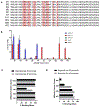
Figure 2:. PD-L1 binding of selected affibody sequences.
A) Sequences of the 10 clones selected for further analysis and binding characterization. Red highlighted amino acids were randomized in the initial library. Blue bolded amino acids were spontaneous mutations arising in the non-randomized regions of the affibody scaffold. B) Sequences were expressed as mRNA-affibody fusions and panned against immobilized hPD-L1, mPD-L1, hPD-L2 or Her2. An asterisk under the affibody indicates it was not assessed for binding to hPD-L2 or Her2 due to poor hPD-L1 binding. C) The clinical PD-L1 binding antibody Tecentriq® (atezolizumab) was used in a blocking assay to determine if selected affibody clones bound at the hPD-L1:PD-1 interface. In almost all clones, hPD-L1 binding was completely blocked relative to the no affibody control (% block binding). D) A similar experiment was carried out with a dual mouse/human PD-L1 binding antibody blocking assay to determine if mPD-L1 selected affibodies bind at the mPD-L1/PD-1 interface.
Figure 3:
Figure 3:. M1 affibody blocks PD-1:PD-L1 signaling in cell culture.
M1 affibody and control Her2 affibody were incubated with Jurkat T-cells expressing PD-1and CHO cells expressing hPD-L1. Blocking the PD-1:PD-L1 signaling axis in T-cells increases bioluminescence. The M1 affibody blocks this interaction with an IC50 of 2 μM (0.7 to 6 μM for 95% confidence interval). The Her2 affibody was used for background normalization.
Figure 4:
Figure 4:. Binding of the M1 Affibody to immobilized PD-L1.
A) Binding of AlexaFlour488nm and B) SulfoCy5 dye-labeled affibodies (M1 and Her2 affibody control) to streptavidin immobilized hPD-L1, mPD-L1, or Her2. The binding is measured as the % of resin-associated fluorescence relative to the total input fluorescence. These experiments demonstrate that the dye-labeled affibodies bind selectively to their immobilized targets. The M1 affibody bound to both C) mPD-L1 and D) hPD-L1 in a concentration-dependent fashion (KD= 47 (38 to 58) nM and 89 (68 to 120) nM, respectively) but not to HER2 or PD-L2. E) As expected, the control HER2 affibody bound only to immobilized Her2 (KD= 11 (7.3 to 16) nM). KD values are provided along with the 95% confidence interval in parentheses (GraphPad Prism).
Figure 5:
Figure 5:. Surface plasmon resonance (SPR) analysis of binding kinetics of M1 Affibody.
Two-fold serial dilutions of M1 affibody were flowed over a biotin-tethered PD-L1 sensor surface on a streptavidin chip. A) SPR sensorgrams for the binding of N-methylmaleimide (NMM)-capped affibody to human PD-L1 surface (~ 700 RU) are shown in black and data were fit to a 1:1 Langmuir binding model (red overlay line). The derived dissociation constant KD was calculated from the rate constants (KD = kd/ka). B) M1 Affibody and mouse PD-L1 (~850 RU) binding was fitted to a two-state model (blue overlay line) in order to describe the complex interaction observed. The apparent dissociation constant KD value was estimated using the binding rate constants (KD = kd1/ ka1), while the conformational change rates (kd2 = 6.94 × 10⁻³ 1/s and ka2 = 1.91 × 10⁻² 1/s) were omitted (see materials and methods). C) The average binding response (RU) at/close to steady state (110-113 s) as a function of affibody concentration is shown.
Figure 6:
Figure 6:. Affibody binding to CHO cells by flow cytometry.
A) M1-AF488nm affibody binding to CHO cells expressing hPD-L1 and normalized to CHO parental cells (KD = 95.5 (81.7 to 111) nM) . B) M1-AF488nm affibody binding to CHO cells expressing mPD-L1 and normalized to CHO parent cells (KD=587 (458 to 751) nM) . C) M1-AF488nm affibody binding to hPD-L2 and normalized to CHO parent cells; no binding was observed. D) HER2-AF488nm affibody binding to SKBr3 (Her2 overexpressing breast cancer cell line) and hPD-L1 expressing CHO cells lines. HER2 affibody bound strongly to SKBr3 cells (KD = 7.12 (6.11 to 8.30) nM) but not to hPD-L1 expressing CHO cells. KD values are provided along with the 95% confidence interval in parentheses (GraphPad Prism)
Figure 7:
Figure 7:. M1 affibody uptake of hPD-L1 expressing tumors.
A) Following IP pre-injection with either atezolizumab (ATZ) or control IgG1 antibody, mice with EL4 lymphoma subcutaneous flank tumors expressing hPD-L1 were imaged on the IVIS® Spectrum In vivo Imaging System to establish background signal arising from autofluorescence. Mice were then injected with either 5 nmol M1-Cy5 affibody or HER2-Cy5 affibody (negative control) and imaged 1 and 4 hours later. Mice were sacrificed to expose tumor and organ uptake (T is tumor, H is heart, L is lungs). Affibody uptake in side-facing tumors B) and ex vivo resected tumors C) was quantified by placing regions of interest (ROI) over the tumor and measuring radiant efficiency. Two-way ANOVA with multiple comparisons show M1 affibody has statistically higher uptake than control HER2 in tumors over all time points and states. D) Lysed and processed tumor tissue evaluated for Cy5 presence show increased amounts of fluorescence (pM Cy5) with M1 affibody in both conditions versus the Her2 affibody. P value *<0.05, **<0.01,***<0.001, ****<0.0001.

References

    1. Vaddepally RK; Kharel P; Pandey R; Garje R; Chandra AB, Review of Indications of FDA-Approved Immune Checkpoint Inhibitors per NCCN Guidelines with the Level of Evidence. Cancers (Basel) 2020, 12 (3). - PMC - PubMed
    1. Alsaab HO; Sau S; Alzhrani R; Tatiparti K; Bhise K; Kashaw SK; Iyer AK, PD-1 and PD-L1 Checkpoint Signaling Inhibition for Cancer Immunotherapy: Mechanism, Combinations, and Clinical Outcome. Front Pharmacol 2017, 8, 561. - PMC - PubMed
    1. De Silva P; Aiello M; Gu-Trantien C; Migliori E; Willard-Gallo K; Solinas C, Targeting CTLA-4 in cancer: Is it the ideal companion for PD-1 blockade immunotherapy combinations? Int J Cancer 2021, 149 (1), 31–41. - PubMed
    1. Thallinger C; Fureder T; Preusser M; Heller G; Mullauer L; Holler C; Prosch H; Frank N; Swierzewski R; Berger W, et al., Review of cancer treatment with immune checkpoint inhibitors : Current concepts, expectations, limitations and pitfalls. Wien Klin Wochenschr 2018, 130 (3-4), 85–91. - PMC - PubMed
    1. Teng F; Meng X; Kong L; Yu J, Progress and challenges of predictive biomarkers of anti PD-1/PD-L1 immunotherapy: A systematic review. Cancer Lett 2018, 414, 166–173. - PubMed

Publication types