A Multimodal Approach to Quantify Chondrocyte Viability for Airway Tissue Engineering
- PMID: 35612419
- PMCID: PMC9691794
- DOI: 10.1002/lary.30206
A Multimodal Approach to Quantify Chondrocyte Viability for Airway Tissue Engineering
Abstract
Objectives/hypothesis: Partially decellularized tracheal scaffolds have emerged as a potential solution for long-segment tracheal defects. These grafts have exhibited regenerative capacity and the preservation of native mechanical properties resulting from the elimination of all highly immunogenic cell types while sparing weakly immunogenic cartilage. With partial decellularization, new considerations must be made about the viability of preserved chondrocytes. In this study, we propose a multimodal approach for quantifying chondrocyte viability for airway tissue engineering.
Methods: Tracheal segments (5 mm) were harvested from C57BL/6 mice, and immediately stored in phosphate-buffered saline at -20°C (PBS-20) or biobanked via cryopreservation. Stored and control (fresh) tracheal grafts were implanted as syngeneic tracheal grafts (STG) for 3 months. STG was scanned with micro-computed tomography (μCT) in vivo. STG subjected to different conditions (fresh, PBS-20, or biobanked) were characterized with live/dead assay, terminal deoxynucleotidyl transferase dUTP nick end labeling (TUNEL), and von Kossa staining.
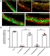
Results: Live/dead assay detected higher chondrocyte viability in biobanked conditions compared to PBS-20. TUNEL staining indicated that storage conditions did not alter the proportion of apoptotic cells. Biobanking exhibited a lower calcification area than PBS-20 in 3-month post-implanted grafts. Higher radiographic density (Hounsfield units) measured by μCT correlated with more calcification within the tracheal cartilage.
Conclusions: We propose a strategy to assess chondrocyte viability that integrates with vivo imaging and histologic techniques, leveraging their respective strengths and weaknesses. These techniques will support the rational design of partially decellularized tracheal scaffolds.
Level of evidence: N/A Laryngoscope, 133:512-520, 2023.
Keywords: Regenerative medicine; biobanking; chondrocyte viability; tissue engineering; tracheal replacement.
© 2022 The Authors. The Laryngoscope published by Wiley Periodicals LLC on behalf of The American Laryngological, Rhinological and Otological Society, Inc.
Conflict of interest statement
Conflict of Interest Statement
We have no conflicts of interest to disclose.
Figures


Similar articles
-
Assessing the Impact of Partial Decellularization on Tracheal Chondrocytes and Extracellular Matrix in Airway Reconstruction.Otolaryngol Head Neck Surg. 2025 Jun;172(6):2026-2037. doi: 10.1002/ohn.1211. Epub 2025 Mar 21. Otolaryngol Head Neck Surg. 2025. PMID: 40114536 Free PMC article.
-
Optimization of Chondrocyte Viability in Partially Decellularized Tracheal Grafts.Otolaryngol Head Neck Surg. 2023 Nov;169(5):1241-1246. doi: 10.1002/ohn.404. Epub 2023 Jun 14. Otolaryngol Head Neck Surg. 2023. PMID: 37313949 Free PMC article.
-
Long-Term Chondrocyte Retention in Partially Decellularized Tracheal Grafts.Otolaryngol Head Neck Surg. 2024 Jan;170(1):239-244. doi: 10.1002/ohn.409. Epub 2023 Jun 27. Otolaryngol Head Neck Surg. 2024. PMID: 37365963 Free PMC article.
-
Decellularized tracheal scaffolds in tracheal reconstruction: An evaluation of different techniques.J Appl Biomater Funct Mater. 2021 Jan-Dec;19:22808000211064948. doi: 10.1177/22808000211064948. J Appl Biomater Funct Mater. 2021. PMID: 34903089 Review.
-
Mechanical Evaluation of Tracheal Grafts on Different Scales.Artif Organs. 2018 May;42(5):476-483. doi: 10.1111/aor.13063. Epub 2017 Dec 11. Artif Organs. 2018. PMID: 29226358 Review.
Cited by
-
Assessing the Biocompatibility and Regeneration of Electrospun-Nanofiber Composite Tracheal Grafts.Laryngoscope. 2024 Mar;134(3):1155-1162. doi: 10.1002/lary.30955. Epub 2023 Aug 14. Laryngoscope. 2024. PMID: 37578209 Free PMC article.
-
Partial Decellularization Retains Cartilage Immune Privilege in Tissue Engineered Tracheal Grafts.Laryngoscope. 2025 Sep;135(9):3232-3239. doi: 10.1002/lary.32227. Epub 2025 May 5. Laryngoscope. 2025. PMID: 40323179
-
Regeneration of tracheal neotissue in partially decellularized scaffolds.NPJ Regen Med. 2023 Jul 12;8(1):35. doi: 10.1038/s41536-023-00312-4. NPJ Regen Med. 2023. PMID: 37438368 Free PMC article.
-
Assessing the Impact of Partial Decellularization on Tracheal Chondrocytes and Extracellular Matrix in Airway Reconstruction.Otolaryngol Head Neck Surg. 2025 Jun;172(6):2026-2037. doi: 10.1002/ohn.1211. Epub 2025 Mar 21. Otolaryngol Head Neck Surg. 2025. PMID: 40114536 Free PMC article.
-
Partial decellularization eliminates immunogenicity in tracheal allografts.Bioeng Transl Med. 2023 Apr 21;8(5):e10525. doi: 10.1002/btm2.10525. eCollection 2023 Sep. Bioeng Transl Med. 2023. PMID: 37693070 Free PMC article.
References
-
- Grillo HC. Surgery of the trachea and bronchi. PMPH USA, 2004.
-
- Kolb F, Simon F, Gaudin R et al. 4-Year follow-up in a child with a total autologous tracheal replacement. N Engl J Med 2018; 378:1355–1357. - PubMed
-
- Delaere P, Vranckx J, Verleden G, De Leyn P, Van Raemdonck D. Tracheal allotransplantation after withdrawal of immunosuppressive therapy. New England Journal of Medicine 2010; 362:138–145. - PubMed
Publication types
MeSH terms
Grants and funding
LinkOut - more resources
Full Text Sources
Medical
Research Materials

