. 2022 Dec;11(1):1604-1620.
doi: 10.1080/22221751.2022.2082888.

Identification and characterization of key residues in Zika virus envelope protein for virus assembly and entry

Affiliations

Identification and characterization of key residues in Zika virus envelope protein for virus assembly and entry

Xiao Ma et al. Emerg Microbes Infect. 2022 Dec.

Abstract

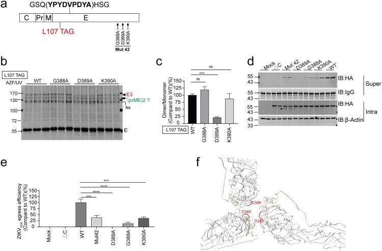
Zika virus (ZIKV), a family member in the Flavivirus genus, has re-emerged as a global public health concern. The envelope (E) proteins of flaviviruses play a dual role in viral assembly and entry. To identify the key residues of E in virus entry, we generated a ZIKV trans-complemented particle (ZIKVTCP) system, in which a subgenomic reporter replicon was packaged by trans-complementation with expression of CprME. We performed mutagenesis studies of the loop regions that protrude from the surface of the virion in the E ectodomains (DI, DII, DIII). Most mutated ZIKVTCPs exhibited deficient egress. Mutations in DII and in the hinge region of DI and DIII affected prM expression. With a bioorthogonal system, photocrosslinking experiments identified crosslinked intracellular E trimers and demonstrated that egress-deficient mutants in DIII impaired E trimerization. Of these mutants, an E-trimerization-dead mutation D389A that nears the E-E interface between two neighbouring spikes in the immature virion completely abolished viral egress. Several mutations abolished ZIKVTCPs' entry, without severely affecting viral egress. Further virus binding experiments demonstrated a deficiency of the mutated ZIKVTCPs in virus attachment. Strikingly, synthesized peptide containing residues of two mutants (268-273aa in DII) could bind to host cells and significantly compete for viral attachment and interfere with viral infection, suggesting an important role of these resides in virus entry. Our findings uncovered the requirement for DIII mediated-E trimerization in viral egress, and discovered a key residue group in DII that participates in virus entry.

Keywords: Zika virus (ZIKV); envelope protein; flavivirus; glycoprotein; mutagenesis; transcomplemented particle system; viral egress; viral entry.


Conflict of interest statement

No potential conflict of interest was reported by the author(s).

Figures

Figure 1.
Figure 1.
Generation and characterization of ZIKV trans-complemented particles (ZIKVTCPs). (a) Replication of ZIKV subgenomic replicon. Upper panel, schematic of ZIKV subgenomic. C25, coding sequence of the first 25-amino acids of capsid. sGluc, secreted Gaussia luciferase. 2A, FMDV 2A peptide. BSD, blasticidin resistance gene. NS1.sig, signal peptide of NS1. HDVr, HDV ribozyme. Bottom panel, replication of sgZIKV-sGluc and sgZIKV-sGluc-GNN with NS5 lethal mutation. The in vitro-transcribed RNAs were transfected into Vero cells. Supernatants were harvested at various time points and the luciferase activities were determined. Mean values ± SDs are shown (n = 3). (b) Generation of the sgZIKV-sGluc stable cell line. SgZIKV-sGluc RNAs were transfected into Vero cells and grown in medium with 5 μg/ml basticidin, and after 15 passages, the cell lysates were analyzed by western blotting with the indicated antibodies. The values to the left of the blots are molecular sizes in kilodaltons. (c and d) Generation of ZIKVTCP. (c) Schematic of the experiment design. Plasmids expressing ZIKV CprM-E or prM-E were transfected into Vero-sgZIKV-sGluc cells. After 96 h, the conditioned medium was harvested and used to infect naïve Vero cells. (d) About 7.5 × 104 Vero cells in the 48-well plate format were infected with 80 μl condition medium for 1 day. Then cells were washed thrice with PBS and fresh media was added. The supernatants were collected at various time points post infection and the luciferase activity in the supernatants was measured. Mean values ± SDs are shown (n = 3). (e and f) Inhibition of ZIKVTCP infection by Bafilomycin and NHCl4. Vero cells were infected with ZIKVTCP in the presence of Bafilomycin (e) and NHCl4 (f) at various concentrations as described in d. At 1-day post infection, cells were washed with PBS and fresh media was added. Supernatants were collected at 1-day and 4-day post infection and luciferase activity were determined. The relative luciferase activity at 4-day post infection relative to 1-day post infection was plotted. Mean values ± SDs are shown (n = 3). Statistical analysis was performed between the treated groups and the control-treated (0) group (***P < 0.001; two-tailed, unpaired t-test). (g) Infection of ZIKVTCPHA. ZIKVTCPHA with HA-tagged E protein (upper panel) was generated as described in (c) by transfecting plasmid expressing CprM-EHA. ZIKVTCPs were used to infect naïve Vero cells as described in (c). The supernatants were harvested at the indicated time points and luciferase activates were measured and plotted. Mean values ± SDs are shown (n = 3).
Figure 2.
Figure 2.
Assembly and infectivity of ZIKVTCP bearing E mutants. (a) Structure of the mature ZIKV virion (PDB, 5iz7). The Loop regions (arrows) selected in this study and the schematic of ZIKV E domains are shown. (b) Schematic of ZIKV E mutagenesis. Three-amino acid mutations (Ala-Ala-Ala) (Red stars) are introduced in the indicated regions. The HA tag is inserted in the DI as indicated. (c) Schematic of the experimental design for D to F. Plasmids expressing CprM-EHA (WT), prM-E (ΔC) and CprM-EHA-mutants were transfected into Vero-sgZIKV-sGluc cells, respectively. At 4 days post transfection, cell lysates (Intra) were harvested for Western blotting (WB) analysis. ZIKVTCPs in the medium were captured by anti-HA beads (super) and analyzed by Western blotting. Alternatively, condition media were harvested and used to infect naïve Vero cells. At various time points, the luciferase activities in the supernatants of the infected cells were determined. (d) Cell lysates (Intra) and captured ZIKVTCPs (Super) were analyzed by western blotting with the indicated antibodies. Representative pictures of Mut 1 to 8 are shown. The values to the left of the blots are molecular sizes in kilodaltons. (e) Summary of the Relative level of EHA secreted into the supernatants to that in the intracellular cell lysates. Relative levels of EHA egress of each mutants were calculated as (captured supernatant HA/Supernatant IgG)/ (Intracellular HA/Intracellular Actin) and further normalized to the WT. Data combined from two independent experiments are shown (mean ± SEM, n = 6). (f) Conditioned media containing ZIKVTCPs were used to infect naïve Vero cells. The Gluc luciferase activities in the supernatants of the infected cells were determined at the indicated time points post infection. Mean values ± SDs are shown (n = 3).
Figure 3.
Figure 3.
Visualization of E trimer by a bioorthogonal system. (a) Left: Structure of ZIKV immature virion (PDB, 6LNU). Four representative trimeric E-prM spikes are shown. Right: The E-prM interfaces within a spike and the E-E interface between two neighbouring spikes and the residues in the interfaces are shown. The residues selected in this study are indicated in red. (b) Alignment of the amino acids from different ZIKV strain and DENV within the interfaces. (c) Localizations of the selected residues (in red) in the interfaces in the structures. The prM in the immature virion is indicated. (d) The sgZIKV-sGluc-Vero cell line was cotransfected with plasmid pSVB.Yam, pcDNA.RS, and plasmids expressing CprM-EHA with the TAG stop codon (red dots) introduced at the indicated residues. After UV-crosslinking, the cells were subjected to immunoprecipitation (IP) with anti-HA beads and the immunoprecipitated proteins were analyzed by Western blotting. The arrows indicate the E trimers (E3) with the expected molecular weight of 165.5 KD, the putative dimeric prME with the expected molecular weight of 148KD and the putative prME with the expected molecular weight of 74KD. The asterisks indicate the E monomers. Ns, nonspecific bands; Us, unspecified bands. The values to the left of the blots are molecular sizes in kilodaltons. (e) The abundances of the E trimers in (d) were quantified and the intensities of the E trimer were calculated as the ratio of E trimer/ E monomer. Mean values ± SD are shown (n = 3). (f) Summary of the results of the photo crosslinking results of all the E mutants. Mean values ± SD are shown (n = 3).
Figure 4.
Figure 4.
Impact of E mutations on E trimerization. (a) Schematic of HA-tagged E mutants with the TAG stop codon introduced at L107. (b) SgZIKV-sGluc-Vero cells were cotransfected with the plasmid pSVB.Yam, pcDNA.RS and the E mutant plasmids. After UV-crosslinking, the cell lysates were subjected to immunoprecipitation (IP) with anti-HA beads. Proteins were analyzed by Western blotting with anti-HA antibody. Representative Western blot of some mutants showing data from triplicate wells are shown. The values to the left of the blots are molecular sizes in kilodaltons. The E trimers (E3) and the putative dimeric prME are indicated. Ns, nonspecific bands. (c) The abundances of the E trimers in (b) were quantified. The relative intensities of the E trimers formed by the mutants were calculated (E trimer/ E monomer) and further normalized to WT. Mean values ± SDs are shown (n = 3). Statistical analysis was performed between WT and the mutants. (ns, not significant; *P < 0.05; **P < 0.01; ***P < 0.001; ****P < 0.0001; two-tailed, unpaired t-test). (d) Visualization of the equivalent residues (in red) of E mutants (Mut 20, Mut 22, Mut 27, Mut 37, Mut 40 and Mut 42) that impair E trimerization in mature virion (PDB, 5iz7) and immature virion (PDB, 6LNU) structures.
Figure 5.
Figure 5.
D389 is essential for E trimerization. (a) Schematic of HA-tagged E mutants with TAG stop codon introduced at L107. (b) SgZIKV-sGluc-Vero cells were cotransfected with the plasmid pSVB.Yam, pcDNA.RS and the E mutant plasmids. After UV-crosslinking, the cell lysates were subjected to immunoprecipitation (IP) with anti-HA beads. Proteins were analyzed by Western blotting with anti-HA antibody. The E trimers (E3) and the putative dimeric prME are indicated. Ns, nonspecific bands. The values to the left of the blots are molecular sizes in kilodaltons. (c) The abundances of the E trimers in (b) were quantified, and the relative intensities of the E trimers were calculated (E trimer/ E monomer) and then further normalized to WT. Mean values ± SDs are shown (n = 3). Statistical analysis was performed between L107 TAG-WT and the variants as indicated (ns, not significant; ***P < 0.001; two-tailed, unpaired t-test). (d) SgZIKV-sGluc-Vero cells were transfected with plasmids expressing CprM-EHA (WT), prM-EHA (ΔC) and CprM-EHA mutants as indicated. At 4-day post transfection, ZIKVTCPs in the supernatants (super) were captured by anti-HA beads and analyzed by Western blotting with anti-HA antibody. The cell lysates (Intra) were analyzed by Western blotting with the indicated antibodies. Representative Western blot with duplicated samples is shown. The values to the left of the blots are molecular sizes in kilodaltons. The arrow indicates a cleaved form of E. (e) The egress efficiency of ZIKVTCPs in (d) were calculated as (captured supernatant HA/Supernatant IgG)/(Intracellular HA/Intracellular Actin). Data combined from two independent experiments are shown (mean ± SEM, n = 4). Statistical analysis was performed between WT and the mutants as indicated (ns, not significant; ***P < 0.001; ****P < 0.0001; two-tailed, unpaired t-test). (f) Visualization of the equivalent residues (in red) of D389 in the immature virion structure (PDB, 6LNU).
Figure 6.
Figure 6.
Identification of residues important for viral entry. (a, b) Concentration of ZIKVTCP. (a) Schematic of ZIKVTCP concentration. Plasmids expressing HA-tagged E mutants Mut 9 (A268/L269A/E270A), Mut 10 (A271/E272A/M273A), Mut 36 (E366A/N367A/S368A) and Mut 37 (K369A/M370A/M371A) were transfected into sgZIKV-sGluc-Vero cells, respectively. ZIKVTCPs in the supernatants were concentrated by ultracentrifugation through 20% sucrose cushion. ZIKVTCPs were normalized to C7.EHA by Western blotting analysis of viral E proteins. (b) Western blotting analysis of ZIKVTCPs by anti-HA antibody (upper panel). A representative Western blot of triplicate samples is shown. Relative abundances of E proteins of each ZIKVTCP to ZIKVTCP-WT were calculated (bottom panel). Mean values ± SDs are shown (n = 3). The values to the left of the blots are molecular sizes in kilodaltons. (c–e) Infection of Vero (c), Huh7 (d), A549 (e) by the concentrated ZIKVTCPs in (b). At 1day post infection, the supernatants were removed, and the infected cells were washed and fresh media was added. The luciferase activities in the supernatants were measured at the indicated time points post infection. Mean values ± SDs are shown (n = 3). (f-i) Binding of ZIKVTCPs to Vero cells. (f) Schematic of the experiment design. (g) Vero cells were infected with the normalized ZIKVTCPs and C7.EHA (MOI, 6) at 4°C for 2 h. Then the cells were washed three times with PBS and then fixed for immunostaining with anti-CD44 and anti-HA antibodies and AF488- and Cy3-conjugated 2nd antibody, respectively. The nucleus (blue) is stained by Hoechst 33342. Cells were observed with a confocal microscope. Representative images are shown. Scale bar, 10 μm. (h) The numbers of E-bound cells in (g) were quantified and plotted. Data combined from three independent experiments are shown (mean ± SEM, n = 6). (i) Vero cells were infected with the normalized ZIKVTCPs and C7.EHA (MOI, 6) at 4°C for 2 h. Then the cells were washed six times with PBS. The bound viral RNAs were extracted and quantified by q-PCR. Mean values ± SEMs from three independent experiments are shown (n = 9). Statistical analysis was performed as indicated (**P < 0.01, ***P < 0.001; ****P < 0.0001; two-tailed, unpaired t-test).
Figure 7.
Figure 7.
Binding of ZIKV E-derived peptides to Vero cells. (a) The P1 and P1-Mut sequences and the locations of the equivalent residues (in red) in the structure (PDB, 5i7z) are shown. The amino acids corresponding to Mut 9 and Mut 10 (green) are underlined. (b) Vero cells were incubated with PBS (Mock), FITC-conjugated peptides P1 and P1-Mut at different concentrations for 2 h at 4°C and then fixed and analyzed by flow cytometry as described in material and methods. (c) The P2 and P2-Mut sequences and the locations of the equivalent residues (highlighted) in the structure (PDB, 5i7z) are shown. The amino acids corresponding to Mut 37 (green) are underlined. (d) Vero cells were incubated with PBS (Mock), FITC-conjugated peptides P2 and P2-mut at different concentrations for 2 h at 4°C and then fixed and analyzed by flow cytometry. The SSC (y-axis) and FITC (x-axis) fluorescence intensities of the cells are shown. Gates to indicate binding of FITC-peptides were set on the mock-treated cells. Cell numbers of group 1 (G1) and group 2 (G2) were plotted. Mean values ± SDs are shown (n = 3). Statistical analysis was performed as indicated (**P < 0.01, ***P < 0.001; ****P < 0.0001; two-tailed, unpaired t-test.)
Figure 8.
Figure 8.
ZIKV E-derived peptides compete for ZIKV binding and infection. (a) Schematic of the experiment design. Vero cells were first incubated with various concentrations of P1 or P1-mut peptides as indicated, and then infected with C7-Gluc、C7 or ZIKVTCP for 2 h at 4°C. After washing, the viral RNAs bound to cells were quantified or fresh media was added to the cells and the luciferase activity was determined at 4 days post infection. (b-c) Quantification of viral RNAs bound to cells. The bound C7-Gluc viral RNAs (b) and C7 viral RNAs (c) were extracted and quantified by q-PCR. Mean values ± SDs from two independent experiments are shown are shown (n = 6). Statistical analysis was performed as indicated (ns, not significant, *P < 0.05; ***P < 0.001, two-tailed, unpaired t-test). (d) Luciferase activity of the C7-Gluc infected cells. Mean values ± SDs are shown (n = 3). Statistical analysis was performed between the peptide-treated groups and the un-treated (PBS) group (ns, not significant, **P < 0.01, two-tailed, unpaired t-test). (e) Luciferase activity in the supernatants of the ZIKVTCP-infected cells. Mean values ± SDs are shown (n = 3). Statistical analysis was performed between the peptide-treated groups and the un-treated (PBS) group (ns, not significant, **P < 0.01, two-tailed, unpaired t-test).

Similar articles

Cited by

References

    1. Gould EA, Solomon T.. Pathogenic flaviviruses. Lancet. 2008 Feb 9;371(9611):500–509. - PubMed
    1. Dick GW, Kitchen SF, Haddow AJ, et al. . Isolations and serological specificity. Trans R Soc Trop Med Hyg. 1952 Sep;46(5):509–520. - PubMed
    1. Saiz JC, Vázquez-Calvo Á, Blázquez AB, et al. . Zika virus: the latest newcomer. Front Microbiol. 2016;7:496. - PMC - PubMed
    1. Musso D, Ko AI, Baud D.. Zika virus infection – after the pandemic. N Engl J Med. 2019 Oct 10;381(15):1444–1457. - PubMed
    1. Lindenbach BD, Thiel H-J, Rice CM.. Flaviviridae: the viruses and their relication. In Knipe DM, Howley PM, (eds.), Fields virology, 5th ed. Philadelphia: Lippincott, Williams & Wilkins; 2007Jan 01:1101–1152.

Substances