. 2022 May 25;17(5):e0268842.
doi: 10.1371/journal.pone.0268842. eCollection 2022.

Combined nerve and vascular ultrasound in thoracic outlet syndrome: A sensitive method in identifying the site of neurovascular compression

Affiliations

Combined nerve and vascular ultrasound in thoracic outlet syndrome: A sensitive method in identifying the site of neurovascular compression

Peter Dollinger et al. PLoS One. .

Abstract

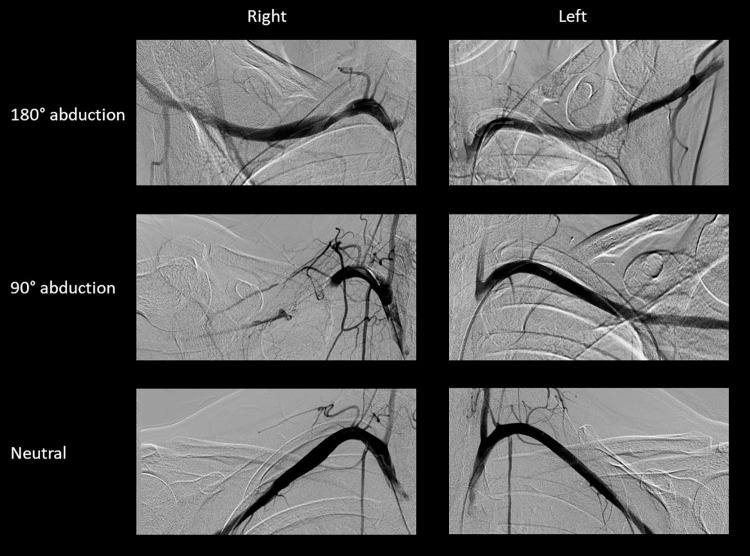
We investigated the diagnostic utility of combined nerve and vascular ultrasound in thoracic outlet syndrome (TOS) in a retrospective cohort study on two sites, involving 167 consecutive patients with the clinical symptoms suggestive of neurogenic and/or vascular TOS, and an age- and sex-matched control group. All patients and control subjects underwent nerve ultrasound of the supraclavicular brachial plexus to look for fibromuscular anomalies / compression of the brachial plexus in the scalenic region, and vascular ultrasound of the infraclavicular subclavian artery with the arm in neutral and abducted position, serving as an indicator for costoclavicular compression of the neurovascular bundle. Based on clinical symptoms, neurogenic TOS (81%) was the most frequent type of TOS, followed by combined neurogenic and arterial TOS (8%). The frequency of abnormal nerve and/or vascular ultrasound findings differed significantly from the control group (P<0.00001). The pooled sensitivity was 48% for nerve ultrasound, 85% for vascular ultrasound, and 94% when combined. Among the findings, the fibromuscular 'wedge-sickle sign', indicating compression of the lower trunk in the scalenic region by a congenital fibromuscular anomaly (e.g. Roos ligaments), showed the highest specificity (100%). A bony 'wedge-sickle sign' was also delineated, where lower trunk compression is caused by the neck of the 1st rib. As implied by the higher sensitivity of vascular ultrasound, the most common site of compression was the costoclavicular space, but multilevel compression was also frequently observed. In summary, combined nerve and vascular ultrasound is a sensitive and reliable method to support the diagnosis of TOS. It can also identify the site(s) of compression, with obvious therapeutic consequences.


Conflict of interest statement

The authors have declared that no competing interests exist.

Figures

Fig 1
Fig 1. The fibromuscular ‘wedge-sickle sign’.
Axial images showing the right brachial plexus in the supraclavicular fossa in patient with neurogenic (supraclavicular) TOS (see also S1 Video). Image B is cephalad relative to image A. In B, note the hyperechoic, ‘wedge’ shaped fibromuscular structure (asterisk) along the caudal medial edge of the middle scalene muscle, indenting (compressing) the lower trunk from the infero-lateral direction, which thus assumes the shape of a ‘sickle’ and becomes swollen and hypoechoic. The fibromuscular structure in this case is a fibrous band connecting the anterior tip of a rudimentary cervical rib (appearing further cephalad as seen in S1 Video) with the first rib (Type 1 Roos ligament). Caudal to the compression (A) the lower trunk is still swollen and hypoechoic, but its shape is round. The middle and upper trunks are normal. Note also the medial insertion of the middle scalene muscle on the 1st rib, forming a V-shaped sling with the anterior scalene muscle and elevating the brachial plexus and the subclavian artery, further restricting space. LT: lower trunk; MD: middle trunk; UP: upper trunk; MS: middle scalene muscle; AS: anterior scalene muscle; Art: subclavian artery.
Fig 2
Fig 2. The bony ‘wedge-sickle sign’.
Axial images showing the right brachial plexus in the supraclavicular fossa in patient with neurogenic (supra- + costoclavicular) TOS. Image B is cephalad relative to image A. In A, note the elevated position of the 1st rib. In B, note that the medial edge of the 1st rib indents the lower trunk from the lateral direction, which assumes a sickle shape and is moderately hypoechoic and swollen. LT: lower trunk; MD: middle trunk; UP: upper trunk; MS: middle scalene muscle; AS: anterior scalene muscle; Art: subclavian artery.
Fig 3
Fig 3. Muscle anomalies in the supraclavicular region.
A. Axial images of the supraclavicular fossa showing the anomalous medial insertion of the middle scalene muscle on the 1st rib in a patient with neurogenic + arterial TOS. Note how the muscle elevates the brachial plexus and has a space-occupying effect. Compare to image A of Fig 2. Further cephalad, a ‘wedge-sickle sign’ is also present (not shown). B. Axial images of the supraclavicular fossa showing an accessory scalenus minimus muscle in a patient with neurogenic + arterial (supra- + costoclavicular) TOS. Note that on the left side, a supernumerary muscle is present between the subclavian artery and the brachial plexus, which has a space-occupying effect. On the right side, the brachial plexus is located immediately adjacent to the artery. Note also the elevated position of the 1st rib. MS: middle scalene muscle; AS: anterior scalene muscle; Art: subclavian artery.
Fig 4
Fig 4. Multilevel compression in neurogenic TOS.
Images of a 20-year-old female patient with neurogenic (supra- + costoclavicular) TOS, demonstrating both supraclavicular ‘wedge-sickle sign’ and costoclavicular impingement. Her complaints started 6 months earlier with nocturnal numbness of the left 4-5th fingers, followed by the development of claw hand, indicating dysfunction of the 4-5th lumbricalis / interosseous (lower trunk innervated) muscles. Apart from the claw hand and minor sensory disturbance in the distribution area of the lower trunk, she had no neurological deficit. Electrophysiological assessment was normal. A. The image on the left is an axial scan of the supraclavicular fossa showing a typical fibromuscular ‘wedge-sickle sign’. Note the sickle-shaped, swollen and hypoechoic lower trunk. The hyperechoic fibromuscular structure in this case is a fibrous band connecting an elongated transverse process of the 7th cervical vertebra (arrows in the middle image) with the 1st rib (Type 2 Roos ligament). The image on the right shows her ‘claw hand’. B and C show the vascular ultrasound (B mode, Colour Doppler and PW mode) of the left subclavian artery in the immediate infraclavicular region in neutral arm position (B) and during arm abduction (C). Note that during arm abduction the artery is narrowed, followed by a poststenotic dilation (‘funnel sign’) as it leaves the costoclavicular space, associated with almost complete occlusion. The patient’s complaints were exacerbated during the manoeuvre. LT: lower trunk; MD: middle trunk; UP: upper trunk; MS: middle scalene muscle; AS: anterior scalene muscle; Clav: clavicula.
Fig 5
Fig 5. Arterial measurements in TOS.
Vascular ultrasound (B mode and PW mode) of the right subclavian artery of a patient with arterial (supra- + costoclavicular) TOS. A shows the artery in the immediate infraclavicular region in neutral arm position, and B during arm abduction. In the neutral position, normal triphasic flow is seen. Note that during arm abduction the artery becomes severely narrowed (arrow, ‘funnel sign’) as it leaves the costoclavicular space, associated with a tardus-parvus type postocclusion blood flow, loss of pulse, and severe diffuse pain and numbness of the arm. Clav: clavicula.
Fig 6
Fig 6. Angiography of the subclavian artery in TOS.
Angiography of the subclavian artery of a patient with arterial TOS on the right side. Note the complete occlusion of the right artery when the arm is in 90° abduction, and the stenosis with poststenotic dilation when in 180° abduction.
Fig 7
Fig 7. Type of symptoms and site of compression in TOS.
Note that the most common site of compression was the costoclavicular space, and the most common type was neurogenic TOS. The Y axis denotes the number of patients.

References

    1. Peete RM, Heriksen MD, Anderson TP. Thoracic outlet syndrome: evaluation of a therapeutic exercise program. Mayo Clin Proc 1956; 31:281–287. - PubMed
    1. Gilliatt RW, Le Quesne PM, Logue V, Sumner AJ. Wasting of the hand associated with a cervical rib or band. J Neurol Neurosurg Psychiatry 1970; 33:615–624. doi: 10.1136/jnnp.33.5.615 - DOI - PMC - PubMed
    1. Durham JR, Yao JS, Pearce WH, Nuber GM, McCarthy WJ 3rd. Arterial injuries in the thoracic outlet syndrome. J Vasc Surg 1995; 21:57–69. doi: 10.1016/s0741-5214(95)70244-x - DOI - PubMed
    1. Illig KA, Doyle AJ. A comprehensive review of Paget-Schroetter syndrome. J Vasc Surg 2010; 51:1538–1547. doi: 10.1016/j.jvs.2009.12.022 - DOI - PubMed
    1. Thompson RW in Illig KA, Thompson RW, Freischlag JA, Donahue DM, Jordan SE, Edgelow PI (Editors). Thoracic outlet syndrome. Springer-Verlag; London, 2013. pp.144, 552.

MeSH terms