. 2022 May 27;8(21):eabm5029.
doi: 10.1126/sciadv.abm5029. Epub 2022 May 25.

Actin-binding protein filamin-A drives tau aggregation and contributes to progressive supranuclear palsy pathology

Affiliations

Actin-binding protein filamin-A drives tau aggregation and contributes to progressive supranuclear palsy pathology

Koyo Tsujikawa et al. Sci Adv. .

Abstract

While amyloid-β lies upstream of tau pathology in Alzheimer's disease, key drivers for other tauopathies, including progressive supranuclear palsy (PSP), are largely unknown. Various tau mutations are known to facilitate tau aggregation, but how the nonmutated tau, which most cases with PSP share, increases its propensity to aggregate in neurons and glial cells has remained elusive. Here, we identified genetic variations and protein abundance of filamin-A in the PSP brains without tau mutations. We provided in vivo biochemical evidence that increased filamin-A levels enhance the phosphorylation and insolubility of tau through interacting actin filaments. In addition, reduction of filamin-A corrected aberrant tau levels in the culture cells from PSP cases. Moreover, transgenic mice carrying human filamin-A recapitulated tau pathology in the neurons. Our data highlight that filamin-A promotes tau aggregation, providing a potential mechanism by which filamin-A contributes to PSP pathology.


Figures

Fig. 1.
Fig. 1.. FLNA protein is abundant in the affected neurons and glial cells of PSP brains.
(A) Silver-stained SDS-PAGE gel from sarkosyl-insoluble fractions (P3) of the brains from four cases with PSP (PSP-5, PSP-6, PSP-9, and Twin-B) and two normal control subjects (NL-3 and NL-5). Arrows point to the >250-kDa protein bands specific for PSP and those in PSP-6, PSP-9, and Twin-B excised for nanoLC-MS/MS analyses. (B and C) Immunoblotting (IB) for TBS-extractable fractions (S1) and P3 of the human brains with anti-FLNA antibody. Subjects in (B) include 11 cases with PSP and 5 normal control subjects. Subjects in (C) include 2 normal control subjects (NL-3 and NL-5), Twin-B, 10 with CBD, 10 with AD, 6 with PD, and 5 with DLB. RD4 and RD3 are 4R-tau and 3R-tau isoform–specific antibodies, respectively, and AT8 is a phosphorylated tau antibody. Glyceraldehyde-3-phosphate dehydrogenase (GAPDH) serves as a loading control. Dashed line indicates boundary line between different membranes. (D) Box plots show quantitative comparisons for FLNA to GAPDH ratio in S1 and for FLNA levels in P3. All values are relative to Twin-B. (E) Immunohistochemistry (IHC) for the frontal cortex of PSP-6 shows that the monoclonal anti-FLNA antibody stains TAs (arrow) and the blood vessels (asterisks). Scale bar, 5 μm. (F) Immunofluorescence for the frontal lobes of PSP-6, Twin-B and PSP-9 with anti-FLNA antibody (magenta) and AT8 (green) show colocalization of FLNA and phosphorylated tau in the neurons, oligodendrocytes (oligos), and astrocytes. Anti–glial fibrillary acidic protein (GFAP) antibody (red) was used to identify astrocytes. Arrows show colocalization of AT8 and FLNA. Coarse granular signals (*) indicate autofluorescence of lipofuscin. Scale bars, 10 μm.
Fig. 2.
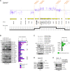
Fig. 2.. Gene duplications of FLNA are identified in the monozygotic twins with PSP.
(A) Enlarged view of microarray data of Twin-A. The copy number gain region includes 16 annotated coding genes, and the copy number changes in a stepwise manner at LCRs within the region. The FLNA gene is duplicated. bp, base pair. (B) Immunoblotting (IB) of the lysates from frontal lobes shows that the levels of proteins coded by 5 (†; FLNA, RPL10, GDI1, FAM3A, and G6PD) of 16 genes within the copy number gains at Xq28 are higher in the both PSP twins (Twin-A and Twin-B) than the mean + SD of five normal controls. (C) IB shows that coexpression of wild-type FLNA (FLNAWT), among the above five proteins, with GFP–4R-tau in HEK293 cells causes significantly higher protein levels of GFP–4R-tau compared to empty (n = 5). (D) IB of the LCLs using total tau antibody TAU-5 shows higher levels of endogenous FLNA and tau in the PSP twins than their siblings (II-1 to II-3). (E) IB of the LCLs from the PSP twins using isoform-specific tau antibodies RD4 and RD3 shows that knockdown of FLNA with siRNAs reduces both 4R-tau and 3R-tau concentrations. Values are presented as means ± SEM. *P < 0.05. Statistics obtained from one-way ANOVA with Tukey-Kramer tests in (C).
Fig. 3.
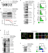
Fig. 3.. FLNA enhances the phosphorylation, protein stability and sarkosyl-insolubility of tau.
(A) Immunoblotting (IB) of the lysates from HEK293 cells shows FLNAWT induces phosphorylation of GFP–4R-tau at the epitopes recognized by the two phosphorylated tau antibodies AT8 (Ser202/Thr205) and PHF-1 (Ser396/Ser404). (B) IB of the lysates from CHX chase assay in HEK293 cells shows that the GFP–4R-tau protein levels at 24 hours are significantly higher when FLNA are coexpressed compared to empty (n = 3). The levels of GFP–4R-tau are normalized to those of GAPDH at 0 hour. (C) IB of the lysates from HEK293 cells shows that overexpression of FLNAWT in HEK293 cells increases GFP–4R-tau concentration in homogenates (Ho), S1, and P3 (n = 3). (D) Immunoprecipitation (IP) assay. More FLNAWT proteins are coimmunoprecipitated with TAU-5 from lysates of HEK293 coexpressing GFP–4R-tau and FLNA. More HSP90, HSP70, HSP40, and ubiquitin are also coimmunoprecipitated. (E) Immunofluorescence shows AT8-immunopositive tau aggregates (blue) in the astrocytes coexpressing GFP–4R-tau (green) and mCherry-tagged FLNA (red) (FLNA+) but not in the astrocytes expressing sole GFP–4R-tau (FLNA−). (F) IB of homogenates from the astrocytes shows that FLNAWT significantly increases the protein levels of GFP–4R-tau with AT8-immunopositive hyperphosphorylation (n = 3). Arrow indicates nonspecific band. Scale bars, 10 μm. Values are presented as means ± SEM. ***P < 0.001, **P < 0.01, and *P < 0.05. Statistics obtained from Tukey-Kramer tests in (C) and Student’s t tests in (F).
Fig. 4.
Fig. 4.. Induction of human FLNA by electroporation enhances GFP–4R-tau protein levels through F-actin in murine brains.
(A) Immunofluorescence for the E18 mouse brains that were electroporated with the indicated plasmids at E14 (n = 15). The electroporation of FLNAWT (red), but not mutant FLNA (FLNAAla39Gly) that abrogates the binding ability to F-actin, results in heterotopia (arrow) and a higher immunoreactivity for GFP–4R-tau (green) compared to empty. The immunoreactivity is normalized to the count of the mCherry-labeled electroporated cells. Scale bars, 100 μm. (B) Immunofluorescence for primary cortical neurons from E15 brain that were electroporated with the indicated plasmids at E14, followed by the treatment of 0.1% dimethyl sulfoxide (DMSO) or 20 nM cytochalasin D (CytoD) (n = 15). Overexpression of FLNAWT (red) with DMSO shows AT8-immunopositive tau aggregates (arrows) and significantly higher area ratio of AT8 (blue) to GFP (green) (% AT8/GFP) on 2 days in vitro (DIV) compared to empty; this phenomenon was attenuated with CytoD treatment. Phalloidin stain (magenta) was used to identify F-actin. Scale bars, 10 μm. (C) Immunofluorescences for the P7 brains that were electroporated with the indicated plasmids at E14. The neurons, oligodendrocytes, and astrocytes in the P7 brains show accumulations of GFP–4R-tau (green) with FLNAWT (red). Anti-GFAP antibody (blue) was used to identify astrocytes. Scale bars, 10 μm. (D) Immunoblotting (IB) of lysates from the P7 brains with the IUE shows that FLNAWT significantly increases the protein levels of GFP–4R-tau compared to empty in S1 and P3 (n = 3). Values are presented as means ± SEM. N.S., not significant. ***P < 0.001, **P < 0.01, and *P < 0.05. Statistics obtained from Tukey-Kramer tests in (A) and (B) and Student’s t tests in (D).
Fig. 5.
Fig. 5.. Stereotaxic injections of AAV-ΔFLNA enhance murine and human tau protein levels in vivo.
(A) Structures of full-length FLNA (FLNAWT) and two shortened FLNA; FLNAABD + Ig1-15+ Ig24 and FLNAABD + Ig9-15+ Ig24 (ΔFLNA). FLNA has N-terminal actin-binding domain (ABD) and 24 immunoglobulin-like internally homologous repeats (Ig1-24). The last C-terminal Ig repeat (Ig24) serves as a dimerization domain. The AAV vectors contain chicken β-actin (CBA) promoter, 6xHis tag, woodchuck hepatitis virus posttranslational regulatory element (WPRE), and SV40 poly(A) signal (SpA). (B) Immunoprecipitation (IP) assay and immunoblotting (IB) with antibody TAU-5 for the lysates from HEK293 cells transfected with the indicated constructs. The protein interaction with FLNA and GFP–4R-tau is shown with the transfections of FLNAABD + Ig1-15+ Ig24 and ΔFLNA. (C) Immunofluorescence using anti-FLNA antibody (red) and total tau antibody K9JA (turquoise) for the coronal sections from 3-month-old (m.o.) wild-type C57BL/6 mouse brains that had been injected stereotaxically at 2 months old with AAV-ΔFLNA or AAV-empty. Asterisks indicate the injected sites. Scale bars, 1 mm. (D) Schema represents the sampling position (#1 to #4) of the AAV-injected brains for IB. Asterisks indicate the injected sites. (E) IB using RD4 and AT8 for homogenates from the AAV-injected brains of the wild-type mice shows that ΔFLNA expression increases protein levels of murine endogenous tau and induces AT8 hyperphosphorylation. (F) Immunofluorescence using anti-6xHis tag antibody (red) and isoform-specific tau antibody RD4 or RD3 (turquoise) for coronal sections from the 3-month-old TAU KO; hT-PAC-N mouse brains that injected stereotaxically at 2 months old with AAV-ΔFLNA or AAV-empty. Scale bars, 10 μm. (G) IB using RD4 and RD3 for homogenates from the injected brains of the TAU KO; hT-PAC-N mice shows that ΔFLNA expression significantly increases protein levels of human 4R-tau and 3R-tau (n = 3). Values are presented as means ± SEM. ***P < 0.001, **P < 0.01, and *P < 0.05. Statistics obtained from Student’s t tests in (G).
Fig. 6.
Fig. 6.. FLNA transgenic mice exhibit endogenous 4R-tau aggregation in the brains.
(A) Immunohistochemistry for the brains of 5-month-old FLNA transgenic mice (FLNA-Tg) shows that anti-FLNA antibody stains in the neurons of cerebral cortex and hippocampus, the oligodendrocytes of corpus callosum, and the astrocytes of hippocampus but not in those of nontransgenic littermate controls (non-Tg). Scale bars, 50 μm. (B) Immunohistochemistry for the brains of 5-month-old FLNA-Tg shows RD4-immunopositive tau deposits in cerebral cortex and hippocampus. Scale bars, 100 μm (left) and 10 μm in the enlarged images (right). (C) Immunofluorescence shows K9JA-immunopositive or AT8-immunopositive tau deposits (turquoise) in the FLNA-stained neurons (red) of FLNA-Tg in the neurons of cortex. Scale bars, 10 μm. (D) Immunoblotting (IB) using anti-FLNA antibody, RD4, and AT8 for the frontal lobes from the 5-month-old FLNA-Tg shows sarkosyl-insoluble phosphorylated 4R-tau (arrowhead) and overexpression of FLNA. (E) Immunofluorescence for primary cortical neurons of FLNA-Tg show a higher area ratio of AT8 (red) to MAP2 (gray) (%AT8/MAP2) and a lower MAP2 immunoreactivity per cells on 6 DIV compared to those of non-Tg, but the knockdown of FLNA with short hairpin RNAs (shRNAs) (shF#1 and shF#2) corrects these findings (n = 12). The control shRNA (shCtr) was used for a negative control. The microtubule-associated protein 2 (MAP2) immunoreactivity is normalized to the count of neurons. Scale bars, 50 μm. Values are presented as means ± SEM. ***P < 0.001, **P < 0.01, and *P < 0.05. Statistics obtained from Tukey-Kramer tests in (E).

References

    1. Boxer A. L., Yu J. T., Golbe L. I., Litvan I., Lang A. E., Höglinger G. U., Advances in progressive supranuclear palsy: New diagnostic criteria, biomarkers, and therapeutic approaches. Lancet Neurol. 16, 552–563 (2017). - PMC - PubMed
    1. Uttl B., Santacruz P., Litvan I., Grafman J., Caregiving in progressive supranuclear palsy. Neurology 51, 1303–1309 (1998). - PubMed
    1. Glasmacher S. A., Leigh P. N., Saha R. A., Predictors of survival in progressive supranuclear palsy and multiple system atrophy: A systematic review and meta-analysis. J. Neurol. Neurosurg. Psychiatry 88, 402–411 (2017). - PubMed
    1. Yoshida M., Astrocytic inclusions in progressive supranuclear palsy and corticobasal degeneration. Neuropathology 34, 555–570 (2014). - PubMed
    1. Ishigaki S., Fujioka Y., Okada Y., Riku Y., Udagawa T., Honda D., Yokoi S., Endo K., Ikenaka K., Takagi S., Iguchi Y., Sahara N., Takashima A., Okano H., Yoshida M., Warita H., Aoki M., Watanabe H., Okado H., Katsuno M., Sobue G., Altered tau isoform ratio caused by loss of FUS and SFPQ function leads to FTLD-like phenotypes. Cell Rep. 18, 1118–1131 (2017). - PubMed