. 2022 May 27;8(21):eabl5032.
doi: 10.1126/sciadv.abl5032. Epub 2022 May 25.

Selective endocytosis of Ca2+-permeable AMPARs by the Alzheimer's disease risk factor CALM bidirectionally controls synaptic plasticity

Affiliations

Selective endocytosis of Ca2+-permeable AMPARs by the Alzheimer's disease risk factor CALM bidirectionally controls synaptic plasticity

Domenico Azarnia Tehran et al. Sci Adv. .

Abstract

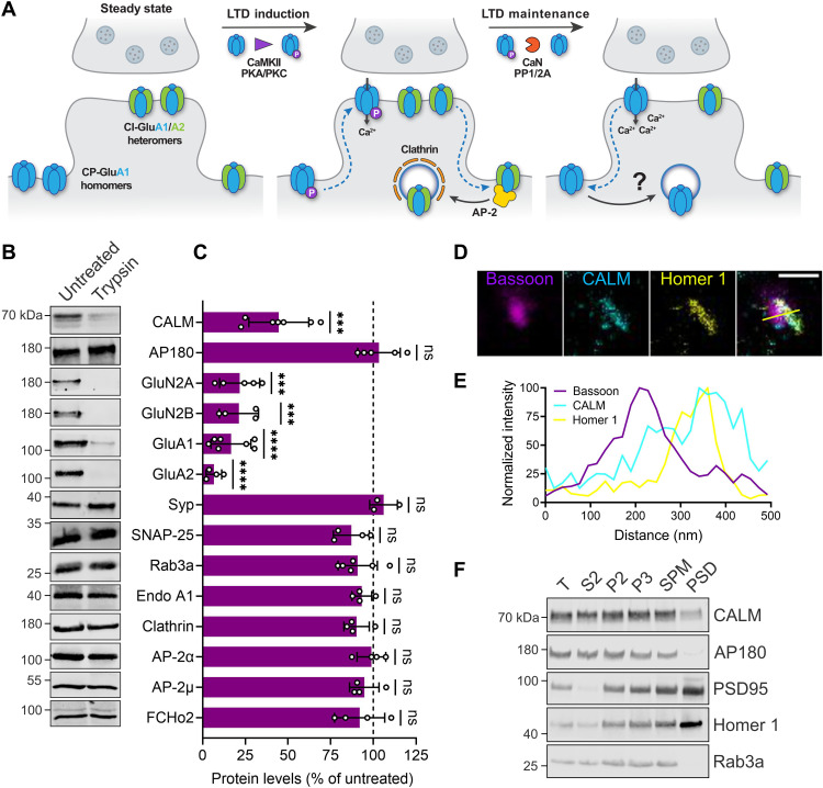
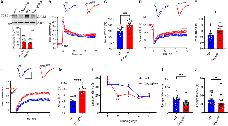
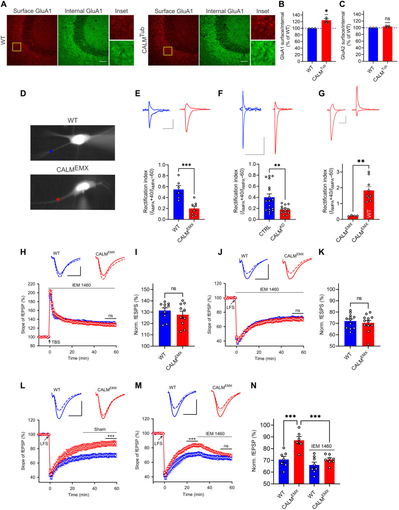
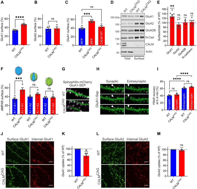
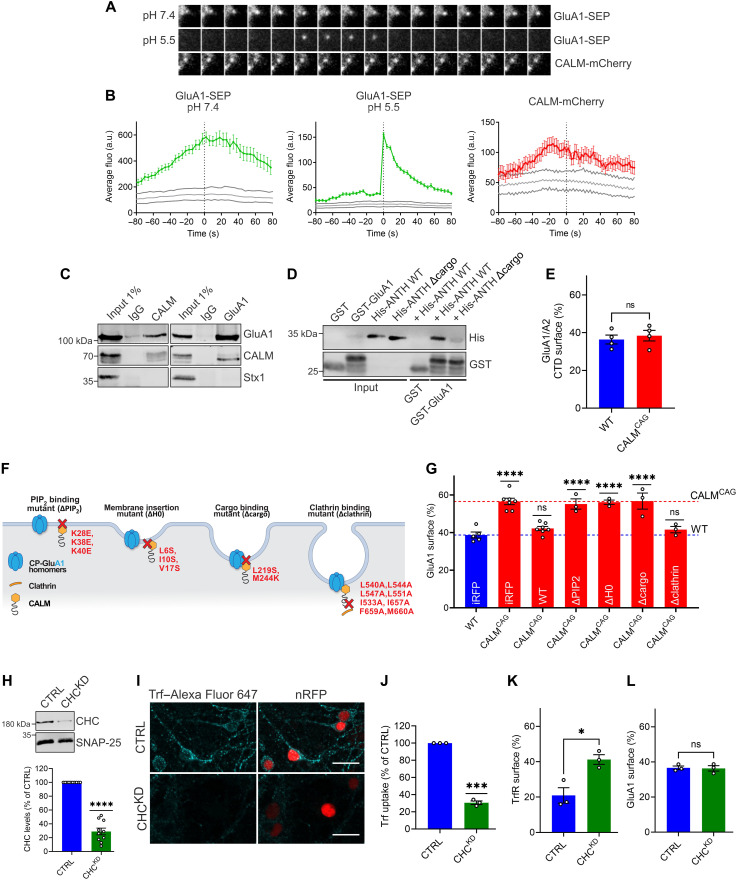
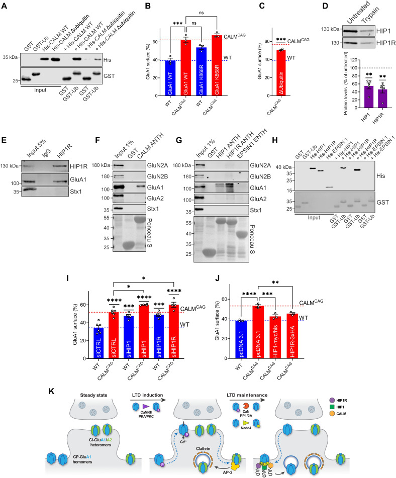
AMPA-type glutamate receptors (AMPARs) mediate fast excitatory neurotransmission, and the plastic modulation of their surface levels determines synaptic strength. AMPARs of different subunit compositions fulfill distinct roles in synaptic long-term potentiation (LTP) and depression (LTD) to enable learning. Largely unknown endocytic mechanisms mediate the subunit-selective regulation of the surface levels of GluA1-homomeric Ca2+-permeable (CP) versus heteromeric Ca2+-impermeable (CI) AMPARs. Here, we report that the Alzheimer's disease risk factor CALM controls the surface levels of CP-AMPARs and thereby reciprocally regulates LTP and LTD in vivo to modulate learning. We show that CALM selectively facilitates the endocytosis of ubiquitinated CP-AMPARs via a mechanism that depends on ubiquitin recognition by its ANTH domain but is independent of clathrin. Our data identify CALM and related ANTH domain-containing proteins as the core endocytic machinery that determines the surface levels of CP-AMPARs to bidirectionally control synaptic plasticity and modulate learning in the mammalian brain.


Figures

Fig. 1.
Fig. 1.. The clathrin adaptor CALM is enriched at postsynaptic endocytic zones where AMPAR endocytosis occurs.
(A) During LTD induction, CP-AMPARs are recruited into synaptic regions, whereas CI-AMPARs are internalized via CME by binding to AP-2. Within a few minutes of LTD progression, the increased Ca2+ concentration activates phosphatases inducing GluA1 dephosphorylation and subsequent CP-AMPAR removal by a still unknown endocytic mechanism. (B and C) Tryptic digest of synaptosomes reveals postsynaptic localization of CALM. (B) Synaptosomes were left untreated or incubated with trypsin and analyzed by immunoblotting. (C) For quantification, trypsin-treated samples were normalized to untreated controls. (D and E) Three-channel time-gated STED confirms predominant postsynaptic localization of CALM. (D) Representative image of wild-type (WT) hippocampal neurons immunolabeled with antibodies against the presynaptic marker Bassoon, the postsynaptic marker Homer 1, and CALM (scale bar, 500 nm). Yellow line indicates position of the normalized fluorescent intensity depicted in (E). (F) The majority of CALM resides outside of the PSD. WT mouse brain homogenates were subjected to subcellular fractionation, and equal protein amounts of total homogenate (T), cytosolic fraction (S2), synaptosomes (P2), synaptosomal membranes (P3), synaptic plasma membranes (SPMs), and PSD were compared by immunoblotting with the indicated markers. Statistics Table 1. ns, not significant.
Fig. 2.
Fig. 2.. CALM bidirectionally controls synaptic plasticity and modulates learning.
(A) Deletion of CALM in KOTub and KOEMX mice. Brain lysates were analyzed by immunoblotting. Protein levels of CALM KO mice were normalized to WT littermates. (B and C) LTP induced by single theta-burst stimulation (TBS) is increased in 2-month-old CALM KOEMX mice. (B) Graph shows normal presynaptic post-tetanic short-term potentiation measured immediately after TBS and increased postsynaptic LTP in CALM KOEMX mice. Top: Representative fEPSPs recorded 0 to 10 min before (solid line) and 50 to 60 min after TBS (dashed line). Scale bar, 0.5 mV and 10 ms. (C) LTP values quantified as percent increase of the responses during the last 10 min. (D to G) LTD is reduced in 2-month-old CALM KOEMX mice and severely impaired in 2-week-old CALM KOEMX mice. (D and F) Top: Representative fEPSPs recorded 0 to 10 min before (solid line) and 50 to 60 min after LFS (dashed line). Scale bars, (D) 1 mV and 10 ms and (F) 0.5 mV and 10 ms. (E and G) LTD values quantified as percent decrease of the responses during the last 10 min. (H to J) CALM KOEMX mice show improved spatial learning. (H) Average escape latency over 5 days of training. Depiction of individual escape latencies for days 2 (I) and 3 (J), where CALM KOEMX mice outperformed controls. Statistics Table 1.
Fig. 3.
Fig. 3.. Surface accumulation of CP-AMPARs in CALM KO mice underlies synaptic plasticity deficits.
(A to C) CALM KO elevates GluA1 surface pools. (A) Brain sections immunostained for GluA1 in the absence/presence of detergent to label surface or internal pool (scale bar, 50 μm). Surface/total ratios of GluA1 (B) or GluA2 (C) normalized to WT. (D) Neurons where AMPAR surface levels at synapses were monitored after glutamate uncaging. (E and F) CALM KO (E) or knockdown (F) leads to decreased AMPAR rectification index (scale bars, 30 pA and 300 ms). (G) Reexpression of CALM in KOEMX slices not only rescues increased inward rectification but also causes outward rectification (scale bars, 30 pA and 300 ms). (H to K) Increased LTP (H) and impaired LTD (J) in CALM-deficient mice are rescued by IEM 1460, applied after induction. Top: fEPSPs recorded 0 to 10 min before (solid) and 50 to 60 min after TBS or LFS (dashed). Scale bars, 0.5 mV and 10 ms (TBS) and 1 mV and 10 ms (LFS). LTP (I) or LTD (K) values quantified as percent change of the responses (last 10 min). (L to N) Impaired LTD in CALM-deficient mice (L) is rescued by IEM 1460, applied 30 min after LTD induction (M). Top: Representative fEPSPs recorded 0 to 10 min before LFS (solid), 20 to 30 min after LFS (before IEM 1460 application; dotted), and 50 to 60 min after LFS (dashed) (scale bars, 1 mV and 10 ms). (N) LTD values quantified as percent change of the responses (last 10 min). Statistics Table 1.
Fig. 4.
Fig. 4.. Loss of neuronal CALM selectively impairs the endocytosis of GluA1 homomers.
(A and B) GluA1 is missorted upon CALM KO. Surface/total ratios of SEP-GluA1 (A) or SEP-GluA2 (B) expressed in WT or KOCAG neurons. (C) Rescue of increased GluA1 surface level in KOCAG neurons by CALM reexpression. Surface/total protein ratios of SEP-GluA1 in neurons transduced with pAAV-iRFP or pAAV-CALM-WT. (D and E) CALM deletion causes increased surface levels of endogenous GluA1. After surface biotinylation of neurons, total and biotinylated proteins were analyzed by immunoblotting (D). Surface/total ratios normalized to WT (E). (F) CALM KO causes the accumulation of GluA1 homomers. Surface/total protein ratios of SEP-tagged GluA1 or GluA2 in WT or KOCAG neurons cotransfected with HA-GluA1 or HA-GluA2 to unravel differences between homomers and heteromers. (G to I) Surface accumulation of GluA1 upon CALM loss not due to altered exocytosis or lateral diffusion. WT and CALM KOCAG neurons coexpressing SEP-GluA1 (green) and dendritic mCherry-spinophilin (magenta) (G) were photobleached at synaptic and extrasynaptic regions (H) (scale bar, 1 μm). Percent fluorescence recovery at 6 to 8 min after beaching (I). (J to M) GluA1 endocytosis is impaired in the absence of CALM. WT and CALM KOCAG neurons live-labeled using antibodies recognizing GluA1 (J) or GluA2 (L) were fixed and immunostained in the absence/presence of detergent to label the surface or internal pool (images: scale bar, 5 μm). GluA1 (K) or GluA2 (M) uptake normalized to WT. Statistics Table 1.
Fig. 5.
Fig. 5.. GluA1 endocytosis is driven by CALM-mediated membrane remodeling and independent of clathrin.
(A and B) CALM-mCherry is present during GluA1 endocytosis events detected with ppH assay at pH 5.5 (A). (B) Average fluorescence intensity aligned to vesicle detection time (frame 0; gray areas, 95% confidence interval of randomized data). (C) Coimmunoprecipitation of endogenous GluA1-CALM complexes from synaptosomal membranes using GluA1- or CALM-specific antibodies. (D) CALM-ANTH domain binds cytosolic GluA1–C terminus. GST–GluA1–C terminus or GST was incubated with His-tagged WT-CALM-ANTH or cargo binding–deficient mutant (Δcargo). Analysis by immunoblotting. (E) Surface levels of GluA1-chimera containing C-terminal domain of GluA2 are unaffected by CALM loss. Neuronal surface/total protein ratios of SEP-GluA1-chimera assessed by acid-base quenching. (F) CALM mutants of (G). (G) CALM’s function in GluA1 endocytosis depends on its PI(4,5)P2 binding and membrane curvature induction but is clathrin independent. SEP-GluA1 surface/total levels in neurons coexpressing iRFP (control), WT-CALM, or CALM-mutant deficient in PI(4,5)P2 binding, membrane insertion (ΔH0), GluA1 binding (Δcargo), or clathrin binding. (H) shRNA-mediated clathrin depletion. Lysates of WT neurons expressing scrambled or anti-CHC shRNA were probed by immunoblotting (clathrin protein levels normalized to SNAP-25). (I and J) Endocytosis of Alexa Fluor 647–transferrin is impaired in clathrin-depleted cells (images: scale bar, 20 μm; nuclear RFP, transduced cells). Values normalized to control cells. (K) Increased transferrin receptor surface pool in clathrin-depleted neurons expressing pHuji-TrfR. (L) Unaltered GluA1 surface pool in clathrin-depleted neurons expressing SEP-GluA1. Statistics Table 1. a.u., arbitrary units.
Fig. 6.
Fig. 6.. Sorting of GluA1 homomers is a general feature of ANTH domain–containing proteins and involves ubiquitin.
(A) CALM-ANTH binds ubiquitin. GST-ubiquitin or GST was incubated with His-WT-CALM-ANTH or its ubiquitin binding–deficient mutant. Analysis by immunoblotting. (B) Ubiquitination and CALM-dependent GluA1 sorting act in the same pathway. Neuronal surface/total protein ratios of SEP-GluA1-WT or its ubiquitination-deficient K868R mutant. (C) Ubiquitin binding–deficient CALM mutant cannot rescue increased GluA1 surface pool of CALM KOCAG neurons. Surface/total SEP-GluA1 protein ratios in KOCAG neurons coexpressing ubiquitin binding–deficient CALM mutant. (D) Tryptic synaptosome digest indicates postsynaptic HIP1/HIP1R localization. Digested synaptosomes were analyzed by immunoblotting (trypsin-treated samples normalized to untreated controls). (E) Coimmunoprecipitation of HIP1R-GluA1 complex from synaptosomal membranes using HIP1R-specific antibodies. (F and G) ANTH domain–containing proteins bind GluA1. GST-ANTH domains of CALM (F), HIP1, HIP1R, or GST-Epsin1-ENTH (G) were incubated with brain lysate. Analysis by immunoblotting. (H) HIP1/HIP1R-ANTH domains, but not Epsin1-ENTH-domain, bind ubiquitin. GST-ubiquitin or GST was incubated with His-tagged proteins. Analysis by immunoblotting. (I) Loss of HIP1 and HIP1R causes GluA1 surface accumulation, especially when combined with CALM deletion. Surface/total SEP-GluA1 protein ratios in neurons treated with control or HIP1- or HIP1R-specific siRNAs. (J) GluA1 surface accumulation upon CALM loss is rescued by overexpression of HIP1 or HIP1R. Surface/total SEP-GluA1 protein ratios in neurons transfected with pcDNA3.1 (=control), HIP1-Myc/His, or HIP1R-3xHA. (K) Endocytic platform of HIP1, HIP1R, and CALM fulfils overlapping cargo-specific role in endocytosis of ubiquitinated GluA1 homomers. Statistics Table 1.

References

    1. Diering G. H., Huganir R. L., The AMPA receptor code of synaptic plasticity. Neuron 100, 314–329 (2018). - PMC - PubMed
    1. Turrigiano G. G., The self-tuning neuron: Synaptic scaling of excitatory synapses. Cell 135, 422–435 (2008). - PMC - PubMed
    1. Groc L., Choquet D., Linking glutamate receptor movements and synapse function. Science 368, (2020). - PubMed
    1. Lee H. K., Takamiya K., He K., Song L., Huganir R. L., Specific roles of AMPA receptor subunit GluR1 (GluA1) phosphorylation sites in regulating synaptic plasticity in the CA1 region of hippocampus. J. Neurophysiol. 103, 479–489 (2010). - PMC - PubMed
    1. Greger I. H., Watson J. F., Cull-Candy S. G., Structural and functional architecture of AMPA-type glutamate receptors and their auxiliary proteins. Neuron 94, 713–730 (2017). - PubMed

Substances