Novel Injury Scoring Tool for Assessing Brain Injury following Neonatal Hypoxia-Ischemia in Mice
- PMID: 35613558
- PMCID: PMC9474591
- DOI: 10.1159/000525244
Novel Injury Scoring Tool for Assessing Brain Injury following Neonatal Hypoxia-Ischemia in Mice
Abstract
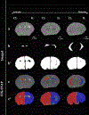
The variability of severity in hypoxia-ischemia (HI)-induced brain injury among research subjects is a major challenge in developmental brain injury research. Our laboratory developed a novel injury scoring tool based on our gross pathological observations during hippocampal extraction. The hippocampi received scores of 0-6 with 0 being no injury and 6 being severe injury post-HI. The hippocampi exposed to sham surgery were grouped as having no injury. We have validated the injury scoring tool with T2-weighted MRI analysis of percent hippocampal/hemispheric tissue loss and cell survival/death markers after exposing the neonatal mice to Vannucci's rodent model of neonatal HI. In addition, we have isolated hippocampal nuclei and quantified the percent good quality nuclei to provide an example of utilization of our novel injury scoring tool. Our novel injury scores correlated significantly with percent hippocampal and hemispheric tissue loss, cell survival/death markers, and percent good quality nuclei. Caspase-3 and Poly (ADP-ribose) polymerase-1 (PARP1) have been implicated in different cell death pathways in response to neonatal HI. Another gene, sirtuin1 (SIRT1), has been demonstrated to have neuroprotective and anti-apoptotic properties. To assess the correlation between the severity of injury and genes involved in cell survival/death, we analyzed caspase-3, PARP1, and SIRT1 mRNA expressions in hippocampi 3 days post-HI and sham surgery, using quantitative reverse transcription polymerase chain reaction. The ipsilateral (IL) hippocampal caspase-3 and SIRT1 mRNA expressions post-HI were significantly higher than sham IL hippocampi and positively correlated with the novel injury scores in both males and females. We detected a statistically significant sex difference in IL hippocampal caspase-3 mRNA expression with comparable injury scores between males and females with higher expression in females.
Keywords: Apoptosis; Brain injury; Caspase-3; Hippocampus; Hypoxia-ischemia; Injury score; MRI; Nuclei integrity; PARP; SIRT; Sirtuin.
© 2022 S. Karger AG, Basel.
Conflict of interest statement
Conflict of Interest Statement
Pelin Cengiz has grant from NIH/NINDS R01 NS111021. Jon E. Levine has grants from NIH R01 DK121559–01, NIH R21 HD102172–01, NIH U24 MH123422–01. The rest of the authors do not have any conflict of interests to declare.
Figures


References
-
- Cowan F, Rutherford M, Groenendaal F, Eken P, Mercuri E, Bydder GM, et al. Origin and timing of brain lesions in term infants with neonatal encephalopathy. Lancet. 2003;361(9359):736–42. - PubMed
-
- Kurinczuk JJ, White-Koning M, Badawi N. Epidemiology of neonatal encephalopathy and hypoxic-ischaemic encephalopathy. Early human development. 2010;86(6):329–38. - PubMed
-
- Ferriero DM. Neonatal brain injury. The New England journal of medicine. 2004;351(19):1985–95. - PubMed
-
- Zhu C, Wang X, Xu F, Bahr BA, Shibata M, Uchiyama Y, et al. The influence of age on apoptotic and other mechanisms of cell death after cerebral hypoxia-ischemia. Cell death and differentiation. 2005;12(2):162–76. - PubMed
-
- Zhu C, Xu F, Wang X, Shibata M, Uchiyama Y, Blomgren K, et al. Different apoptotic mechanisms are activated in male and female brains after neonatal hypoxia-ischaemia. Journal of neurochemistry. 2006;96(4):1016–27. - PubMed
Publication types
MeSH terms
Substances
Grants and funding
LinkOut - more resources
Full Text Sources
Research Materials
Miscellaneous

