. 2022 May 25;13(1):2915.
doi: 10.1038/s41467-022-30576-1.

The structural basis of Cdc7-Dbf4 kinase dependent targeting and phosphorylation of the MCM2-7 double hexamer

Affiliations

The structural basis of Cdc7-Dbf4 kinase dependent targeting and phosphorylation of the MCM2-7 double hexamer

Almutasem Saleh et al. Nat Commun. .

Abstract

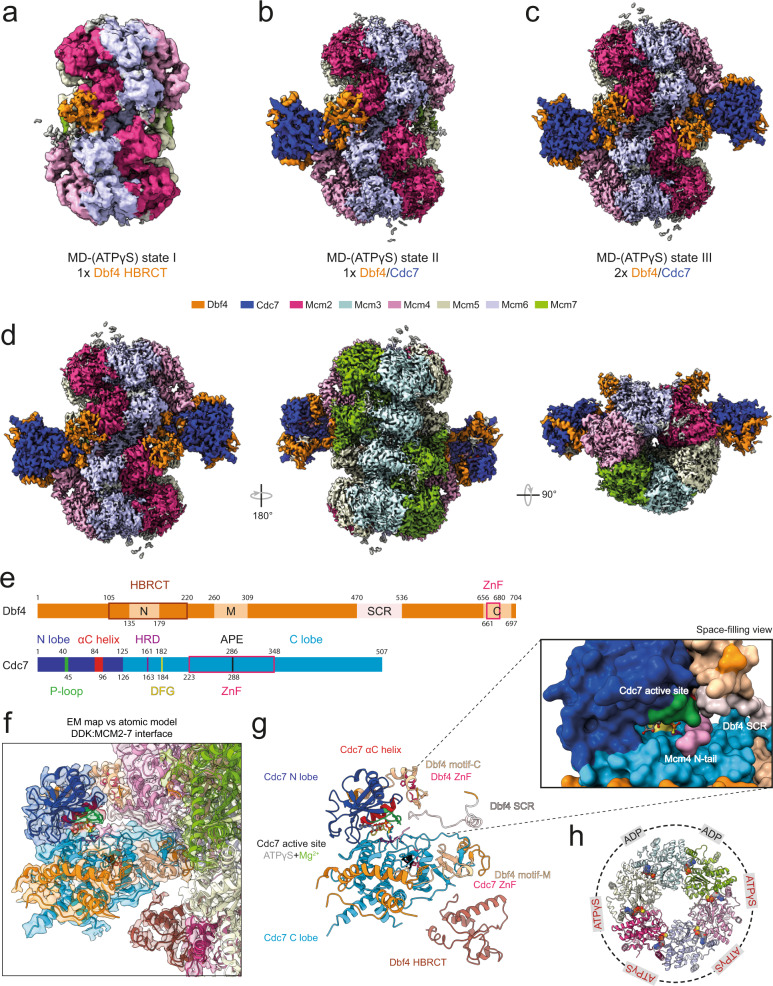
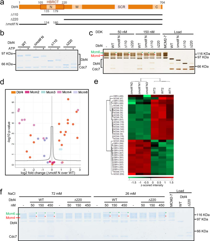
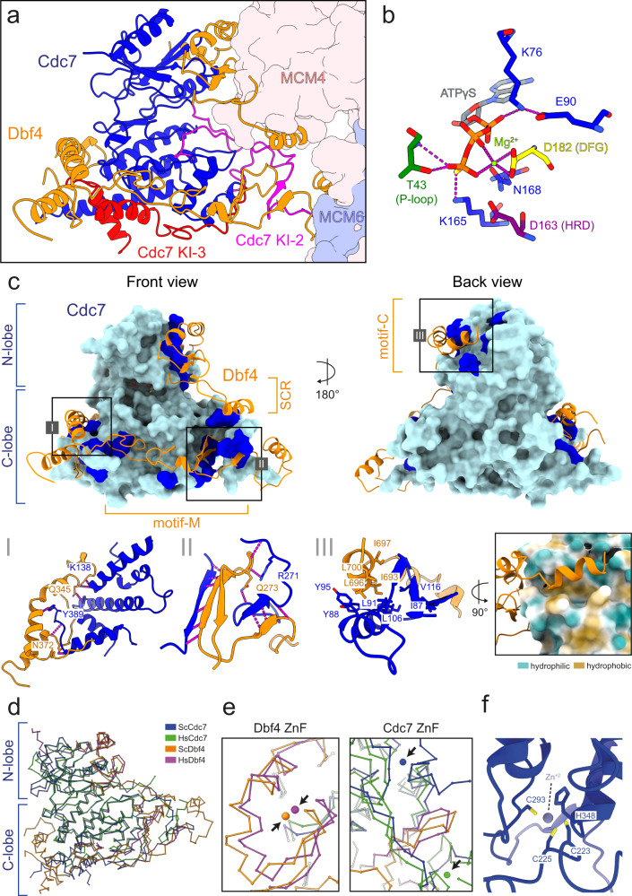
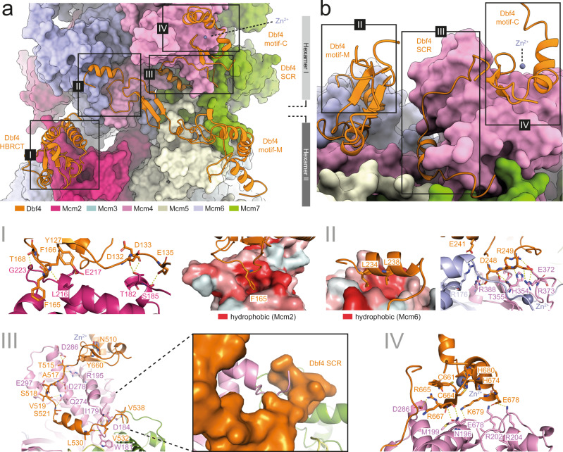
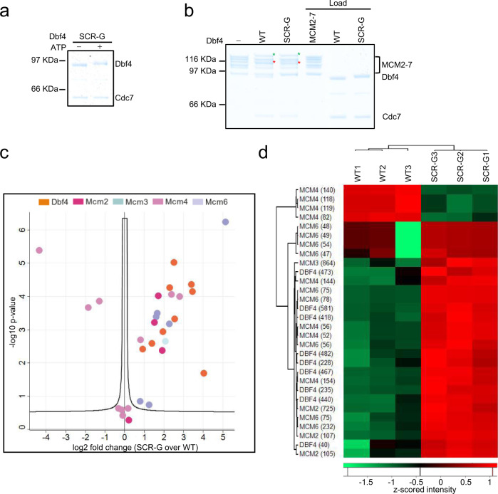
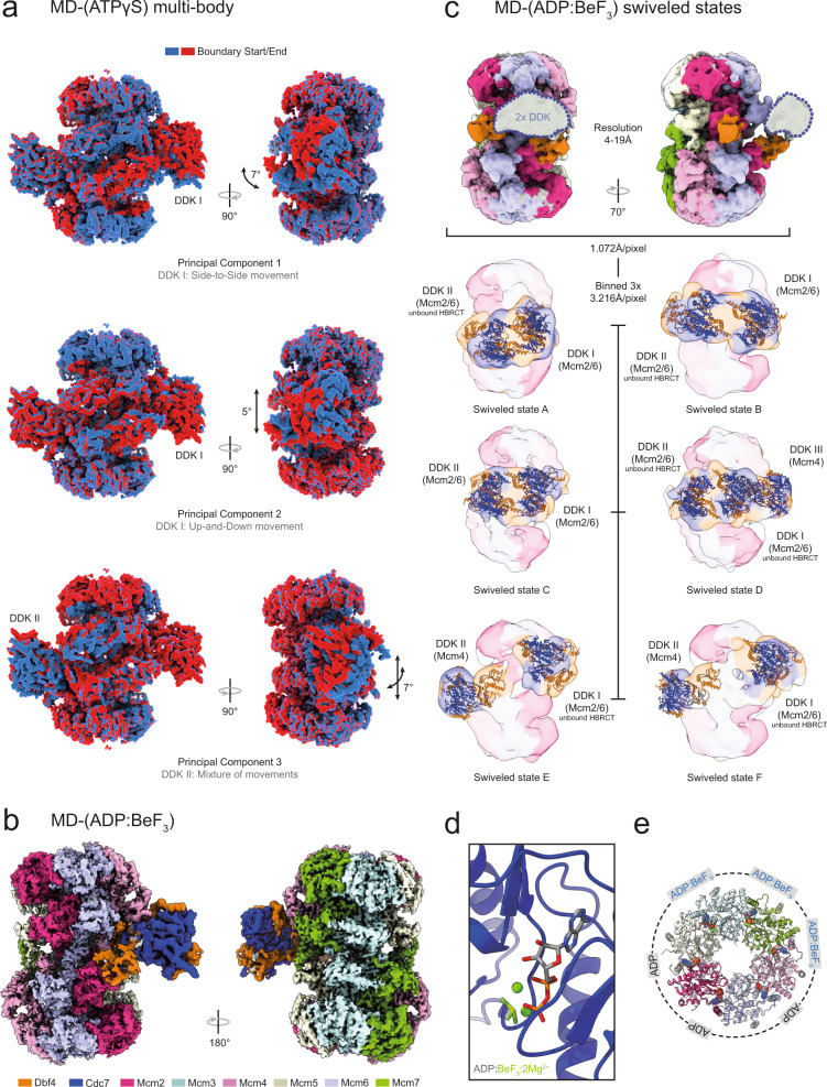
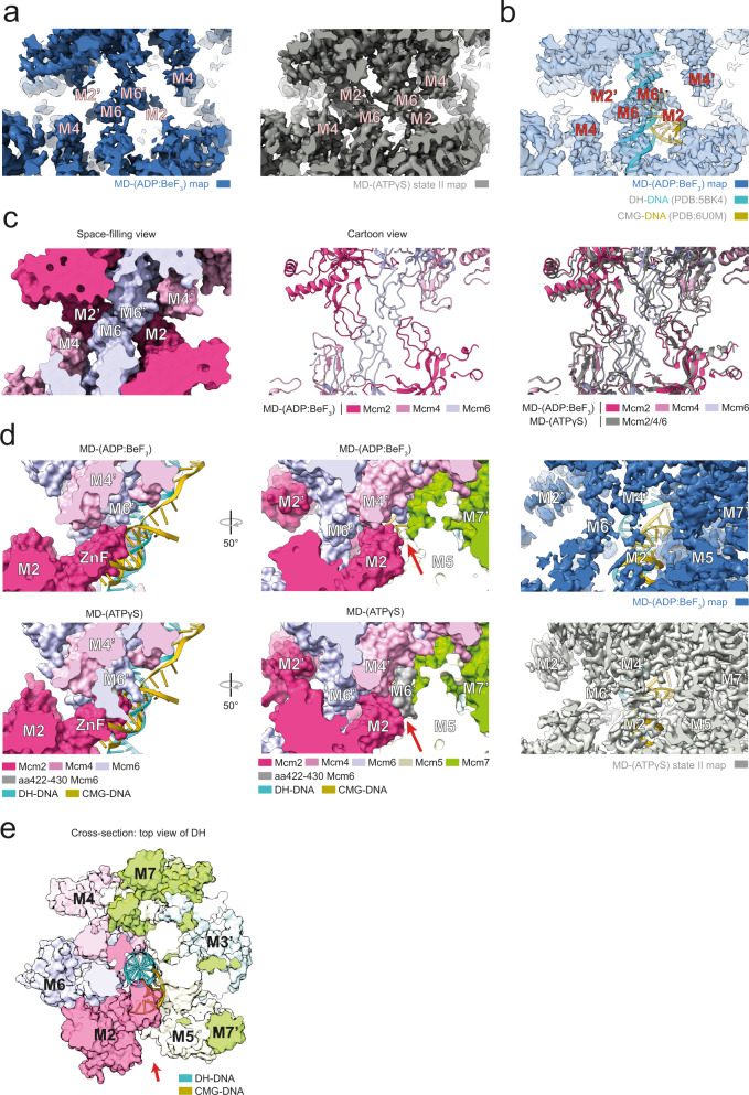
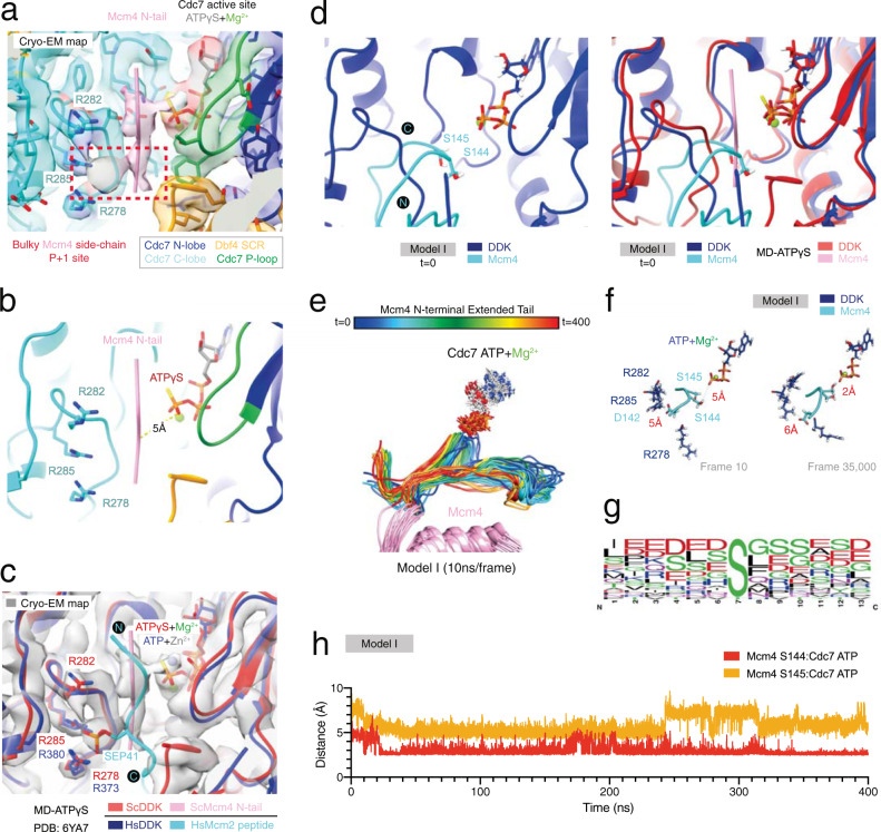
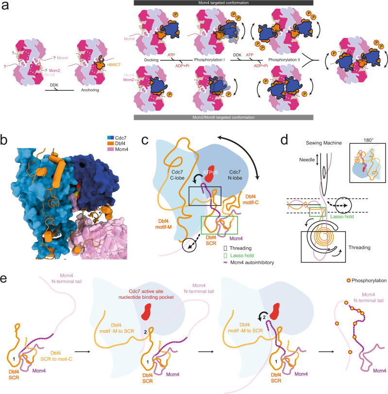
The controlled assembly of replication forks is critical for genome stability. The Dbf4-dependent Cdc7 kinase (DDK) initiates replisome assembly by phosphorylating the MCM2-7 replicative helicase at the N-terminal tails of Mcm2, Mcm4 and Mcm6. At present, it remains poorly understood how DDK docks onto the helicase and how the kinase targets distal Mcm subunits for phosphorylation. Using cryo-electron microscopy and biochemical analysis we discovered that an interaction between the HBRCT domain of Dbf4 with Mcm2 serves as an anchoring point, which supports binding of DDK across the MCM2-7 double-hexamer interface and phosphorylation of Mcm4 on the opposite hexamer. Moreover, a rotation of DDK along its anchoring point allows phosphorylation of Mcm2 and Mcm6. In summary, our work provides fundamental insights into DDK structure, control and selective activation of the MCM2-7 helicase during DNA replication. Importantly, these insights can be exploited for development of novel DDK inhibitors.


Conflict of interest statement

S.S., N.L.C., and M.J.S. are employees of InstaDeep Ltd. The remaining authors declare no competing interests.

Figures

Fig. 1
Fig. 1. Structure of DDK bound to the MCM2-7 double hexamer in the presence of ATPγS.
ad Three different structural states (I-III) derived from the same MD-(ATPγS) cryo-EM data set. a Cryo-EM 3D auto-refined map (see Methods) of MD-(ATPγS) state I. b Composite map (see Methods) of MD-(ATPγS) state II. c, d Composite map (see Methods) of MD-(ATPγS) state III with side and top views. DH at 3.2 Å mean resolution and DDK at 3.6 Å mean resolution. The map density corresponding to each protein subunit component of the complex is coloured according to the key shown. e A schematic diagram illustrating the 2D domain organization the 2D domain organization of Dbf4 and Cdc7. f Comparison of the MD-(ATPγS) atomic model to the cryo-EM map to show the quality of fit. EM map and atomic model are coloured according to key shown in (e). g Same view as (f), but focused only on DDK. The structural features of Cdc7 and Dbf4 are indicated, and a close-up view of the active site is shown. h Overview of the nucleotide occupancy and type in each Mcm subunit within the MD-(ATPγS) complex.
Fig. 2
Fig. 2. The Dbf4 N-terminus is important for DDK dependent phosphorylation of MCM2-7.
a Schematic of Dbf4 N-terminal mutants. The Dbf4 regulatory protein features three unique motifs and a modified domain: motif-N, motif-M and motif-C and α-helix-BRCA1 C-terminal (HBRCT) domain. The substrate coordinating region (SCR) is a newly identified domain. b Analysis and comparison of Dbf4-Cdc7 autophosphorylation ability using Dbf4 wild-type and N-terminal mutants. Similar results were obtained in two independent experiments. c Analysis of Dbf4 N-terminal mutants assessing the interaction of DDK with MCM2-7 DH and MCM2-7 phosphorylation (using 50 and 150 nM DDK concentration). Similar results were obtained in three independent experiments. d Volcano plot comparing the DDK phosphorylation profile of the MCM2-7 DH of WT and Δmotif N Dbf4. Two-sample Student’s t-test carried out with three replicate intensities considered per group. P-values were corrected for multiple comparisons to an FDR of 0.05 (permutation-based FDR). e Volcano plot significant phosphosites visualised using Hierarchical Clustering Analysis (HCA) coupled to a heatmap of z-scored site intensities. f Analysis of WT and Δ220 Dbf4 using different protein and salt concentrations. The results highlight that DDK regions within Δ220 Dbf4 do not support binding to the MCM2-7 DH and only very weakly support MCM2-7 phosphorylation activity. Similar results were obtained in two independent experiments.
Fig. 3
Fig. 3. DDK organization and intra-molecular interactions.
a Front view of DDK bound to the DH. The Cdc7 kinase insert (KI) regions are indicated, KI-2 in magenta and KI-3 in red. b Zoomed view of the Cdc7 active site bound to ATPγS and a Mg2+ ion. The Cdc7 residues surrounding the nucleotide are shown. D182 (DFG motif), T43 (P-loop), and D163 (HRD motif) are coloured in yellow, dark green and purple, respectively and other Cdc7 residues are coloured in blue. c Front and back view of the core of DDK. The surface of Cdc7 is coloured in cyan and regions which contact Dbf4 are highlighted in blue. Zoomed view of the interaction interfaces between: I Dbf4 motif-M and a helical bundle within the Cdc7 C-lobe, II Dbf4 motif-M and Cdc7 KI-3 and III Dbf4 motif-C and a hydrophobic groove on the surface of the Cdc7 N-lobe. ac Cdc7 and Dbf4 are coloured in blue and orange, respectively and hydrogen bond interactions are represented by dotted magenta-coloured lines. d Overall 3D organization of the core of human DDK (PDB:6YA7) and budding yeast DDK (MD-(ATPγS)). Both kinase structures are highly similar and display an active kinase conformation. e Overlay of the zinc finger (ZnF) domains of Dbf4 motif-C and Cdc7 from human and budding yeast. The Dbf4 ZnF is at same 3D position and the Cdc7 ZnF is located at the back of Cdc7 but deviates by 21 Å. f Zoomed view of the budding yeast Cdc7 ZnF and the residues involved in zinc ion coordination.
Fig. 4
Fig. 4. Analysis of the DDK:MCM2-7 interaction interface.
a Cartoon view of Dbf4 and space-filling view of the MCM2-7 DH. Dbf4 forms four independent interaction interfaces along the surface of the MCM-2-7 DH. b Enlarged and rotated view of (a) showing interaction interfaces II–IV. I–IV Detailed view of each of the four Dbf4:Mcm interaction interfaces. The residues involved in making contacts are labelled. The hydrophobic surface shown is coloured according to the Eisenberg hydrophobicity scale.
Fig. 5
Fig. 5. Biochemical analysis of the Dbf4 substrate coordinating region (SCR).
a Dbf4 autophosphorylation analysis of Dbf4 SCR-G. Similar results were obtained in two independent experiments. b The Dbf4 SCR-G mutant was analysed for its interaction of DDK with MCM2-7 DH. 150 nM DDK was used in the reaction. Similar results were obtained in three independent experiments. c Volcano plot comparing WT and SCR-G DDK phosphorylation of the MCM2-7 DH. Two-sample Student’s t-test carried out with three replicate intensities considered per group. P-values were corrected for multiple comparisons to an FDR of 0.05 (permutation-based FDR). d Volcano plot significant phosphosites visualised using HCA coupled to a heatmap of z-scored site intensities.
Fig. 6
Fig. 6. DDK dynamics revealed through multi-body refinement and flexible analysis and alternative MD complex conformations in the presence of ADP:BeF3.
a Flexible analysis of the multi-body refinement of MD-(ATPγS) state III shows distinct movements of DDK relative to the MCM2-7 DH. In addition to DDK movement, the DH C-terminal domain also shows rotational movements. b Side and top views of the cryo-EM composite map (see Methods) of the MD complex in the presence of ADP:BeF3. DH at 3.8 Å mean resolution and DDK at 4.4 Å mean resolution. c The alternative swiveled structural states of the MD complex in the presence of ADP:BeF3. The EM map derived from the unbinned data displayed density large enough to fit two DDK subunits in a swiveled conformation, but the data had to be Fourier binned 3 × 3 to obtain easier to interpret EM maps. The data revealed a range of DDK binding modes, that feature in some cases either or both Mcm2/6 and Mcm4 targeted DDK conformations. The Mcm subunit which is targeted by each DDK is labelled. In some states, the HBRCT domain of Dbf4 is not bound to Mcm2. The local resolution EM maps of the different MD-(ADP:BeF3) swiveled states are shown, coloured according to the key shown in (b). The atomic model of DDK/Dbf4-HBRCT, derived from the map shown in (b), was manually docked into the EM maps. d Zoomed view of the Cdc7 active site bound to ADP:BeF3 and 2 Mg2+ ions. e Overview of the nucleotide occupancy and type in each Mcm subunit within the MD-(ADP:BeF3) complex.
Fig. 7
Fig. 7. Analysis of structural differences within the MD complex in the presence of different nucleotides.
a Zoomed view of the DH central region and comparison between MD-(ADP:BeF3) state I and MD-(ATPγS) state III cryo-EM maps. The MD-(ADP:BeF3) map displays stronger density at the Mcm4/6 zinc finger domains and weaker density at the Mcm2 zinc finger domain compared to MD-(ATPγS). b MD-(ADP:BeF3) state I map and DNA from DH and CMG docked structures. c Comparison between MD-(ADP:BeF3) state I and MD-(ATPγS) state III atomic models. The models show global structural differences at Mcm2/4/6 zinc finger domain regions. d Zoomed view of the Mcm2:Mcm5 interface and comparison between MD-(ADP:BeF3) and MD-(ATPγS) atomic models. A short Mcm6 region (aa422-430) blocks this channel in the MD-(ATPγS) and is disordered in MD-(ADP:BeF3) leaving a 15-30 Å channel opening. e Cross-section of the MD-(ADP:BeF3) atomic model showing the trajectory of DH and CMG DNA between Mcm2:Mcm5. d, e Red arrows indicate channel opening region.
Fig. 8
Fig. 8. Molecular dynamics simulation reveals the binding mode of an extended Mcm4 N-terminal tail substrate at the Cdc7 active site.
a Zoomed view of the MD-(ATPγS) state III atomic model DDK active site and associated cryo-EM density map. The cryo-EM map features density reminiscent of a bulky Mcm4 tail residue side chain at the P + 1 site. This Mcm4 tail unknown residue is also surrounded by Cdc7 R278, R282 and R285. b Simplified view of (a) displaying the distance measurement between the Mcm4 N-terminal tail (backbone CA atom) and Cdc7 ATPγS (O3G atom). c Overlay of the core of human DDK (PDB:6YA7) and budding yeast DDK atomic models. The position of the phospho-serine residue (SEP) in the human Mcm2 peptide matches with the bulky residue side chain density position observed in the MD-(ATPγS) cryo-EM map. d Zoomed view of the DDK active site of Model I (molecular dynamics starting model I, featuring an extended Mcm4 N-terminal tail) and comparison with the MD-(ATPγS) atomic model. e Snapshots of model trajectory, sampled every 10 ns, during a 400 ns GROMACS molecular dynamics simulation. The successive order of the models is represented by rainbow colours (red to blue). The long flexible Mcm4 tail adopts multiple conformations throughout the simulation. f Simplified view of the DDK active site, showing the distance measurements between the nearest DDK Mcm4 target residue (S144, OG atom) and Cdc7 ATPγS (O3G atom) and Mcm4 D142 (CG atom) at the P-2 position and R285 (NE atom). g Sequence logo, generated using WebLogo 3, displaying the frequency (N) of different amino acid residues at atypical human DDK target sites. The residues preceding the target DDK residue feature in a high proportion of cases acidic residues (D/E). h Plot of distance measurement between the Mcm4 N-terminal tail (backbone CA atom) and Cdc7 ATPγS (O3G atom) throughout the entire simulation.
Fig. 9
Fig. 9. Mechanism of DDK substrate localisation and processive phosphorylation of multiple Mcm N-terminal tails.
a Schematic illustration of the different stages of DDK recruitment to the MCM2-7 DH and different modes of binding. b DDK bound to the Mcm4. c Cartoon drawing of (b), with missing structural regions extended as dotted lines. DDK forms a lasso around the most N-terminal resolved region of Mcm4. The missing region between Dbf4 motif-M and Dbf4 SCR traps the Mcm4 flexible tail, encircling it at or close to the Cdc7 active site. d DDK is likened to a sewing machine. The main principle being that Mcm4 gets threaded by DDK, and this process allows the kinase to reach the most C-terminal end of the flexible tail. e Simplified version of (c) showing a step-wise hypothetical mechanism of Mcm4 substrate localisation and processive phosphorylation. The region labelled 1, features a part of Dbf4 SCR which forms a hook (lasso) around the rigid surface of Mcm4. The region labelled 2, features an encircled Mcm4 flexible tail that gets threaded through the kinase. The missing structural regions are represented as dotted lines and resolved regions as solid lines.

References

    1. Jones MJK, et al. Human DDK rescues stalled forks and counteracts checkpoint inhibition at unfired origins to complete DNA replication. Mol. Cell. 2021;81:426–441.e428. doi: 10.1016/j.molcel.2021.01.004. - DOI - PMC - PubMed
    1. Takahashi TS, Basu A, Bermudez V, Hurwitz J, Walter JC. Cdc7-Drf1 kinase links chromosome cohesion to the initiation of DNA replication in Xenopus egg extracts. Genes Dev. 2008;22:1894–1905. doi: 10.1101/gad.1683308. - DOI - PMC - PubMed
    1. Weinreich M, Stillman B. Cdc7p-Dbf4p kinase binds to chromatin during S phase and is regulated by both the APC and the RAD53 checkpoint pathway. EMBO J. 1999;18:5334–5346. doi: 10.1093/emboj/18.19.5334. - DOI - PMC - PubMed
    1. Buck V, White A, Rosamond J. CDC7 protein kinase activity is required for mitosis and meiosis in Saccharomyces cerevisiae. Mol. Gen. Genet. 1991;227:452–457. doi: 10.1007/BF00273937. - DOI - PubMed
    1. Yamada M, Masai H, Bartek J. Regulation and roles of Cdc7 kinase under replication stress. Cell Cycle. 2014;13:1859–1866. doi: 10.4161/cc.29251. - DOI - PMC - PubMed

Publication types

MeSH terms

Substances