

Non-structural proteins of bovine viral diarrhea virus
- PMID: 35614328
- PMCID: PMC9131992
- DOI: 10.1007/s11262-022-01914-8
Non-structural proteins of bovine viral diarrhea virus
Abstract
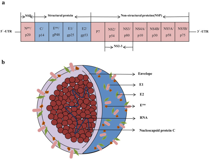
Bovine viral diarrhea virus (BVDV) belongs to the family Flaviviridae genus pestivirus. The viral genome is a single-stranded, positive-sense RNA that encodes four structural proteins (i.e., C, Erns, E1, and E2) and eight non-structural proteins (NSPs) (i.e., Npro, p7, NS2, NS3, NS4A, NS4B, NS5A, and NS5B). Cattle infected with BVDV exhibit a number of different clinical signs including diarrhea, abortion, and other reproductive disorders which have a serious impact on the cattle industry worldwide. Research on BVDV mainly focuses on its structural protein, however, progress in understanding the functions of the NSPs of BVDV has also been made in recent decades. The knowledge gained on the BVDV non-structural proteins is helpful to more fully understand the viral replication process and the molecular mechanism of viral persistent infection. This review focuses on the functions of BVDV NSPs and provides references for the identification of BVDV, the diagnosis and prevention of Bovine viral diarrhea mucosal disease (BVD-MD), and the development of vaccines.
Keywords: Bovine viral diarrhea virus (BVDV); Functions; Nonstructural proteins (NSPs); Vaccine.
© 2022. The Author(s), under exclusive licence to Springer Science+Business Media, LLC, part of Springer Nature.
Conflict of interest statement
The authors declare that they have no competing interests.
Figures


References
-
- Li YM, Liu ZR, Wu YL. Chinese J Vet Sci. 1983;5:113–120.
Publication types
MeSH terms
Substances
Grants and funding
LinkOut - more resources
Full Text Sources
