. 2022 May 9:13:782967.
doi: 10.3389/fneur.2022.782967. eCollection 2022.

SPECT Imaging of Acute Disc Herniation by Targeting Integrin α5β1 in Rat Models

Affiliations

SPECT Imaging of Acute Disc Herniation by Targeting Integrin α5β1 in Rat Models

Jian Guan et al. Front Neurol. .

Abstract

Objective: Traditional morphological imaging of intervertebral disc herniation (IVDH) is challenging in early disease diagnosis. Aiming at the early diagnosis of IVD by non-invasive molecular imaging targeting of integrin α5β1, we performed novel imaging in rats with acute IVDH for the first time.

Methods: Animal models were prepared by conducting an established needle puncture procedure through the normal intervertebral disc (IVD). The disc-injured rats underwent SPECT/CT imaging of the 99mTc-3PisoDGR2 peptide at 1 day to 2 months postinjury. The expression change of integrin α5β1 was determined by anti-integrin α5 and anti-integrin α5β1 immunohistochemistry (IHC). Magnetic resonance imaging (MRI) was performed for comparison during disease progression. The morphological changes of the disc were determined by safranin-O staining.

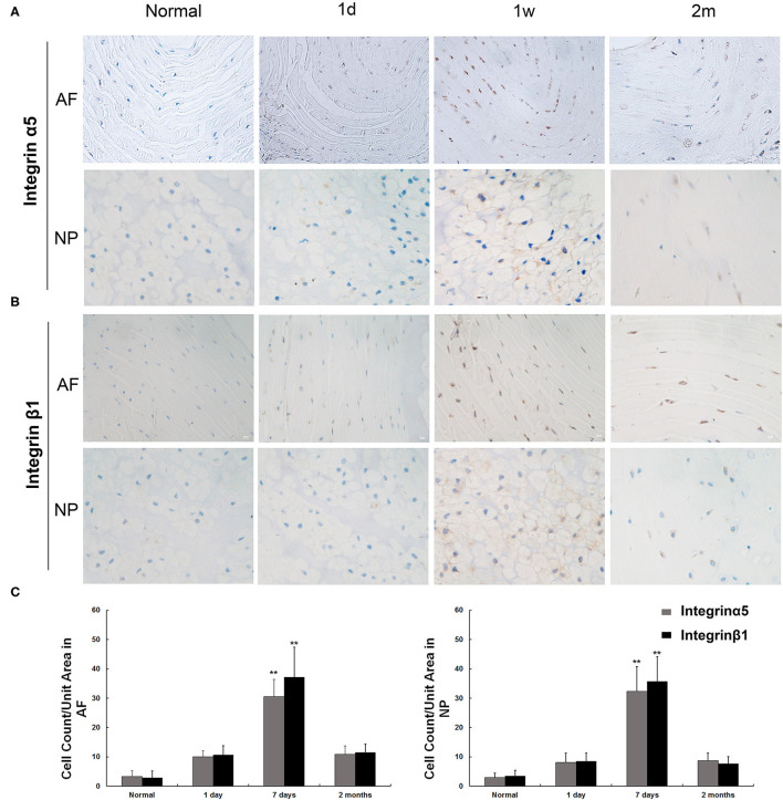
Results: Rats with acute IVDH showed gradually increased disc uptake of 99mTc-3PisoDGR2 from 1 to 7 days posttreatment, which was a significantly higher level than that of the normal disks in degenerative diseases. IHC results showed the expression of integrin α5β1 on the surface of annulus fibrosus (AF) cells and nucleus pulposus (NP) cells, which agreed with the uptake data. MRI showed a progressively decreased T2 density and MRI index throughout the investigation. Hematoxylin and eosin (HE) staining and safranin-O staining revealed a disorganized structure of the IVD as well as loss of proteoglycans after puncture.

Conclusions: The present study demonstrated a good correlation between integrin α5β1 expression and acute disc herniation. The SPECT/CT imaging of 99mTc-3PisoDGR2 targeting integrin α5β1 may diagnose IVDH in an acute phase for early disease management.

Keywords: 99mTc-3PisoDGR2; acute; disc herniation; integrin α5β1; molecular imaging.


Conflict of interest statement

The authors declare that the research was conducted in the absence of any commercial or financial relationships that could be construed as a potential conflict of interest.

Figures

Figure 1
Figure 1
(A) Schematic diagram of the model of disc herniation induced in rats by conducting an established needle puncture procedure. (B) Experimental flowchart. SPECT and MRI were performed 1 day, 1 week, 1 month, and 2 months postinjury. Histology was performed 1 day, 1 week, and 2 months postinjury (IHC, immunohistochemistry; HRMR, high-resolution MRI; HE, hematoxylin and eosin; SO, safranin-O staining).
Figure 2
Figure 2
Representative SPECT images in the caudal spine at 1 day, 7 days, 1 month, and 2 months postinjury. (A) Radioactive signal accumulation was revealed in the punctured disc area of the injured disc at 1 day postinjury. The radioactivity peaked at 1 week and remained at a higher level until 2 months postinjury. A small amount of radioactivity was found in normal disks. (B) The radioactivity of the disc injury region in vivo was plotted against the post probe injection time. **p < 0.01 compared to injured disks at 1 day, 1 month, and 2 months. (C) To validate the specificity of 99mTc-3PisoDGR2 for integrin α5β1, we conducted 99mTc-RGD imaging in a rat model of disc herniation. 99mTc-RGD is the specific probe for integrin αvβ3, which was intravenously injected into the injury group (n = 2) on day 7 postinjury. The experimental process was the same as before. Almost no focal signal was observed in the corresponding region relative to the normal disc of the RGD group.
Figure 3
Figure 3
Immunohistochemical staining of integrin α5 and integrin β1 in nucleus pulposus (NP) and annulus fibrosus (AF) cells. (A) The expression of integrin α5 was observed in normal AF and NP cells. The increased expression of integrin α5 appeared as early as 1 day postinjury, and the peak appeared at 7 days postinjury and remained visible until 60 days. (B) The expression of integrin α5 was observed in normal AF and NP cells. The increased expression of integrin β1 appeared as early as 1 day postinjury, and the peak appeared at 7 days postinjury and remained visible until 60 days. (C) Integrin α5- and β1-positive cells per unit tissue volume were plotted at various time points (1 day, 7 days, and 2 months) after rat disc puncture. **p < 0.01 compared to normal disc and injured disc at 1 day and 2 months.
Figure 4
Figure 4
(A) Representative serial T2-weighted midsagittal images of the caudal disks 1 day, 7 days, 1 month, and 2 months postinjury. (B) Quantitative analysis of T2 density. (C) Quantitative analysis of the MRI index.
Figure 5
Figure 5
HE and safranin-O staining of injured disks at various time points (1 day, 7 days, 1 month, and 2 months). (A) HE staining revealed that the needle puncture induced structural changes (white arrow) in the tissue of the herniated IVD. (B) Safranin-O staining demonstrated that after stab perturbation, the positively stained areas became much smaller (black arrow), indicating decreased proteoglycan content. HE, hematoxylin and eosin.

Similar articles

References

    1. Vialle LR, Vialle EN, Suarez Henao JE, Giraldo G. Lumbar disc herniation. Rev Bras Ortop. (2010) 45:17–22. 10.1590/S0102-36162010000100004 - DOI - PMC - PubMed
    1. Long DM, BenDebba M, Torgerson WS, Boyd RJ, Dawson EG, Hardy RW, et al. . Persistent back pain and sciatica in the United States: patient characteristics. J Spinal Disord. (1996) 9:40–58. 10.1097/00002517-199602000-00007 - DOI - PubMed
    1. Garrido E. Lumbar disc herniation in the pediatric patient. Neurosurg Clin N Am. (1993) 4:149–52. 10.1016/S1042-3680(18)30616-8 - DOI - PubMed
    1. Mayer HM, Mellerowicz H, Dihlmann SW. Endoscopic discectomy in pediatric and juvenile lumbar disc herniations. J Pediatr Orthop B. (1996) 5:39–43. 10.1097/01202412-199605010-00008 - DOI - PubMed
    1. Suthar P, Patel R, Mehta C, Patel N. MRI evaluation of lumbar disc degenerative disease. J Clin Diagn Res. (2015) 9:TC04–9. 10.7860/JCDR/2015/11927.5761 - DOI - PMC - PubMed