. 2022 May 9:16:883584.
doi: 10.3389/fnins.2022.883584. eCollection 2022.

Navigated Intraoperative 3D Ultrasound in Glioblastoma Surgery: Analysis of Imaging Features and Impact on Extent of Resection

Affiliations

Navigated Intraoperative 3D Ultrasound in Glioblastoma Surgery: Analysis of Imaging Features and Impact on Extent of Resection

Benjamin Saß et al. Front Neurosci. .

Abstract

Background: Neuronavigation is routinely used in glioblastoma surgery, but its accuracy decreases during the operative procedure due to brain shift, which can be addressed utilizing intraoperative imaging. Intraoperative ultrasound (iUS) is widely available, offers excellent live imaging, and can be fully integrated into modern navigational systems. Here, we analyze the imaging features of navigated i3D US and its impact on the extent of resection (EOR) in glioblastoma surgery.

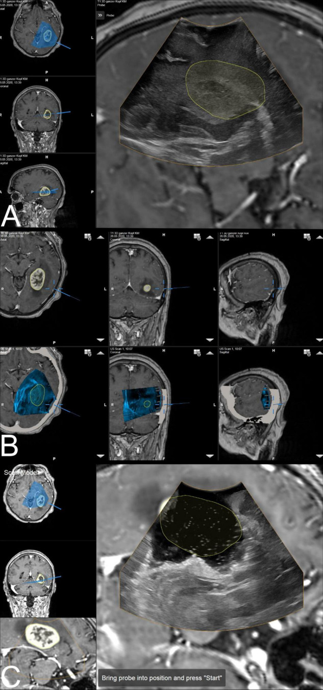
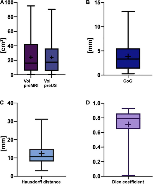
Methods: Datasets of 31 glioblastoma resection procedures were evaluated. Patient registration was established using intraoperative computed tomography (iCT). Pre-operative MRI (pre-MRI) and pre-resectional ultrasound (pre-US) datasets were compared regarding segmented tumor volume, spatial overlap (Dice coefficient), the Euclidean distance of the geometric center of gravity (CoG), and the Hausdorff distance. Post-resectional ultrasound (post-US) and post-operative MRI (post-MRI) tumor volumes were analyzed and categorized into subtotal resection (STR) or gross total resection (GTR) cases.

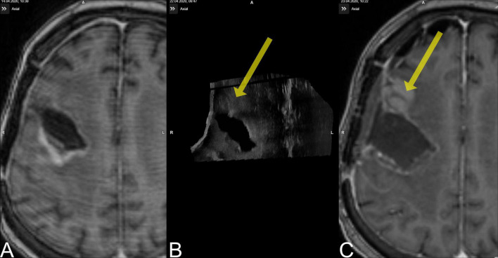
Results: The mean patient age was 59.3 ± 11.9 years. There was no significant difference in pre-resectional segmented tumor volumes (pre-MRI: 24.2 ± 22.3 cm3; pre-US: 24.0 ± 21.8 cm3). The Dice coefficient was 0.71 ± 0.21, the Euclidean distance of the CoG was 3.9 ± 3.0 mm, and the Hausdorff distance was 12.2 ± 6.9 mm. A total of 18 cases were categorized as GTR, 10 cases were concordantly classified as STR on MRI and ultrasound, and 3 cases had to be excluded from post-resectional analysis. In four cases, i3D US triggered further resection.

Conclusion: Navigated i3D US is reliably adjunct in a multimodal navigational setup for glioblastoma resection. Tumor segmentations revealed similar results in i3D US and MRI, demonstrating the capability of i3D US to delineate tumor boundaries. Additionally, i3D US has a positive influence on the EOR, allows live imaging, and depicts brain shift.

Keywords: brain shift; extent of resection; glioblastoma; intraoperative imaging; intraoperative ultrasound; neuronavigation.


Conflict of interest statement

CN and MB are consultants for Brainlab. The remaining authors declare that the research was conducted in the absence of any commercial or financial relationships that could be construed as a potential conflict of interest.

Figures

FIGURE 1
FIGURE 1
The ultrasound application: intraoperative display of i3D US over MRI data. (A) The first intraoperative ultrasound before dural opening is displayed as an overlay. The yellow line shows the tumor borders segmented on pre-MRI, which are not correspondent with i3D US, indicating brain deformation. Corresponding axial, coronal, and sagittal view of pre-MRI data is shown on the left side, including a visualization of localization and orientation of the recent i3D US. (B) After i3D US acquisition, the ultrasound application automatically displays pre-operative imaging (upper row: pre-MRI) and intraoperative imaging (lower row: i3D US as an overlay in pre-MRI) simultaneously in an axial, coronal, and sagittal slice depending on the position of the navigated transducer (cursor). Besides this, the arrows on the right side of each view allow for scrolling through the co-registered i3D US and pre-MRI sets to further explore the data. (C) The second navigated intraoperative ultrasound after resection (post-US) is displayed as an overlay on the pre-MRI. Analogous to (A), the tumor outlines based on the pre-MRI data are visualized in yellow demonstrating the tumor boundaries to be completely within the resection cavity. For comparison of live ultrasound images and pre-MRI data, the scan mode view is chosen. In this case, the left-hand side of the image shows the axial and coronal view, as well as the inline view of the pre-MRI dataset, on which the ultrasound data is superimposed, without the overlay.
FIGURE 2
FIGURE 2
Box-and-whisker plots of pre-resectional data. The lines indicate the medians, boxes extend from the 25th to 75th percentile, the whiskers encompass the range; + indicates the mean. (A) Tumor volumes: Vol pre-MRI 24.2 ± 22.3 cm3 (mean ± SD) and median 16.3 cm3, Vol pre-US 24.0 ± 21.8 cm3 (mean ± SD) and median 17.6 cm3; no significant difference (p = 0.8752, two-tailed Wilcoxon-matched pairs test). (B) Euclidean distance of the center of gravity: 3.9 ± 3.0 mm (mean ± SD), median of 3.3 mm. (C) Hausdorff distance: 12.3 ± 6.9 mm (mean ± SD), median of 10.7 mm. (D) Dice coefficient: 0.71 ± 0.21 (mean ± SD), median of 0.79 (unitless).
FIGURE 3
FIGURE 3
Case no. 9. (A) The pre-MRI (8 days prior to surgery) demonstrated a contrast-enhancing lesion occipital to the resection cavity. (B) The pre-US demonstrated a lesion rostral to the resection cavity (yellow arrow) that was missed during surgery. (C) The new lesion is evident on post-MRI.

Similar articles

Cited by

References

    1. Audette M. A., Siddiqi K., Peters T. M. (eds) (1999). Level-Set Surface Segmentation and Fast Cortical Range Image Tracking for Computing Intrasurgical Deformations. Berlin: Springer Berlin Heidelberg.
    1. Bastos D. C. A., Juvekar P., Tie Y., Jowkar N., Pieper S., Wells W. M., et al. (2021). Challenges and opportunities of intraoperative 3D ultrasound with neuronavigation in relation to intraoperative MRI. Front. Oncol. 11:656519. 10.3389/fonc.2021.656519 - DOI - PMC - PubMed
    1. Brown T. J., Brennan M. C., Li M., Church E. W., Brandmeir N. J., Rakszawski K. L., et al. (2016). Association of the extent of resection with survival in glioblastoma: a systematic review and meta-analysis. JAMA Oncol. 2 1460–1469. 10.1001/jamaoncol.2016.1373 - DOI - PMC - PubMed
    1. Burri S. H., Gondi V., Brown P. D., Mehta M. P. (2018). The evolving role of tumor treating fields in managing glioblastoma: guide for oncologists. Am. J. Clin. Oncol. 41 191–196. 10.1097/COC.0000000000000395 - DOI - PMC - PubMed
    1. Carl B., Bopp M., Sass B., Nimsky C. (2018). Intraoperative computed tomography as reliable navigation registration device in 200 cranial procedures. Acta Neurochir. 160 1681–1689. 10.1007/s00701-018-3641-6 - DOI - PubMed

LinkOut - more resources