. 2022 Aug;31(8):1270-1275.
doi: 10.1111/exd.14614. Epub 2022 Jun 13.

Trichothiodystrophy hair shafts display distinct ultrastructural features

Affiliations

Trichothiodystrophy hair shafts display distinct ultrastructural features

Angeliki-Diotima Ioannidis et al. Exp Dermatol. 2022 Aug.

Abstract

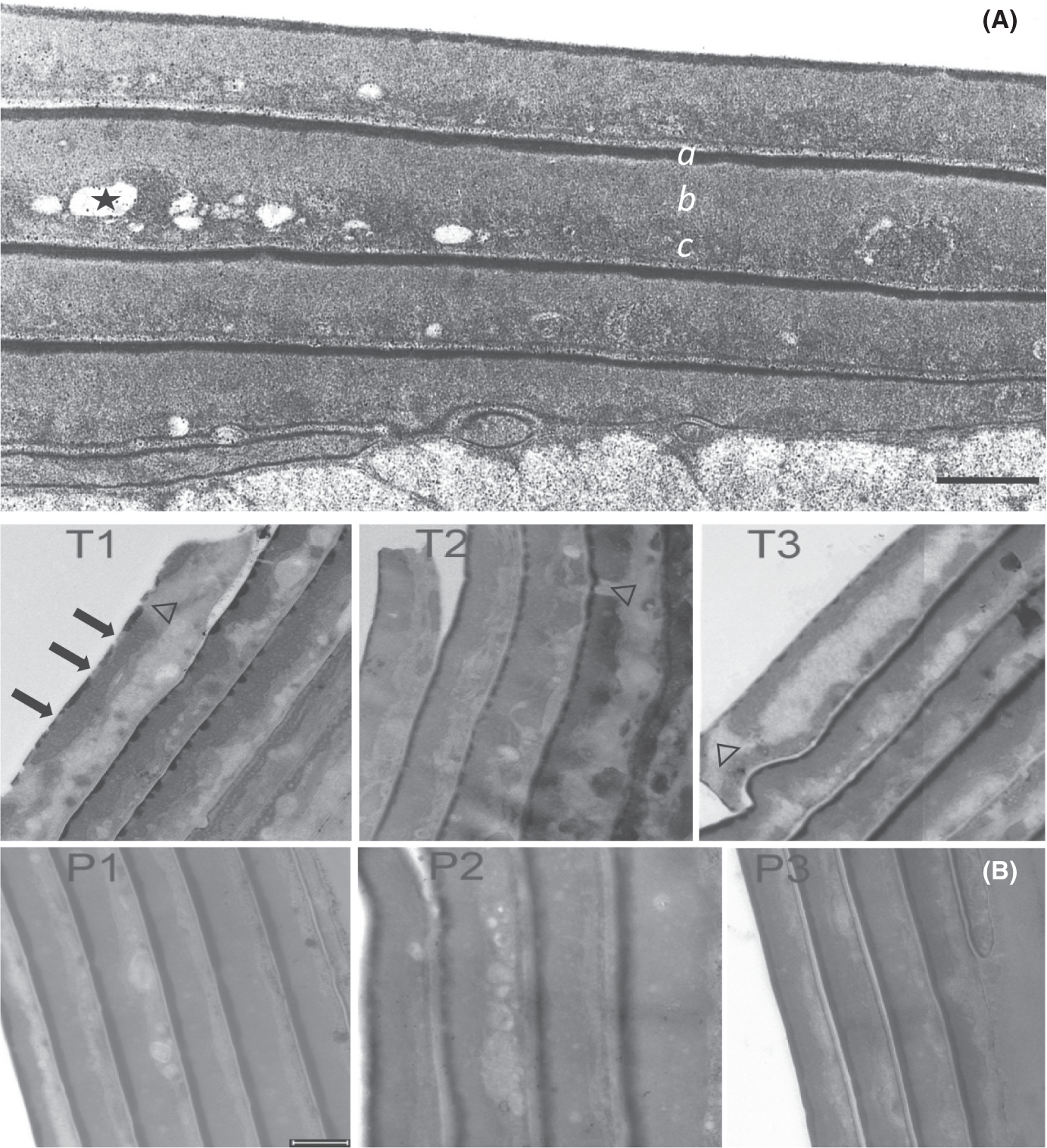
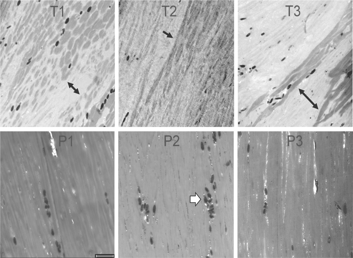
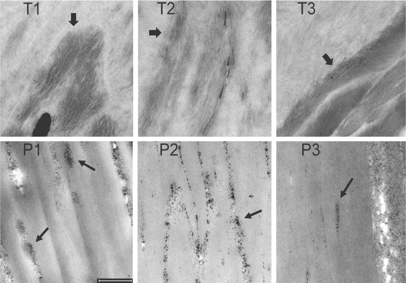
Hair shafts from three trichothiodystrophy (TTD) patients with mutations in the ERCC2 (XPD) gene were examined by transmission electron microscopy. TTD is a rare, recessive disorder with mutations in several genes in the DNA repair/transcription pathway, including ERCC2. Unlike previous studies, the hair shafts were examined after relaxation of their structure by partial disulphide bond reduction in the presence of sodium dodecyl sulphate, permitting improved visualization. Compared with hair shafts of normal phenotype, TTD cuticle cells displayed aberrant marginal bands and exocuticle layers. Clusters of cells stained differently (light versus dark) in the cortex of aberrant shafts, and the keratin macrofibrils appeared much shorter in the cytoplasm. Considerable heterogeneity in these properties was evident among samples and even along the length of single hair shafts. The results are consistent with not only a paucity of high sulphur components, such as keratin-associated proteins, but also a profound imbalance in protein content and organization.

Keywords: DNA repair/transcription disease; hair cortex; hair exocuticle; keratin macrofibrils; marginal band; neuroectodermal genodermatosis; transmission electron microscopy.


Conflict of interest statement

CONFLICT OF INTEREST

The authors state that they have no conflict of interest.

Figures

FIGURE 1
FIGURE 1
Transmission electron micrographs. (A) Cuticle of normal hair incubated in 2% SDS – 25 mM DTT at pH 7.8 for 6 h. The shaft was fixed, stained, sectioned and examined by transmission electron microscopy as described. The dark layer on the outer edge of each cell (a) is called the marginal band or A-layer. The fine-grained layer immediately beneath the marginal band is called the exocuticle (b). The coarse-grained layer at the bottom edge of the cell is the endocuticle (c), in which remnants of intracellular organelles frequently appear embedded, some of which are extracted by the harsh detergent treatment (one indicated by *). (B) Hair shaft cuticle from TTD patients (T1, T2, T3), their respective parents (P1, P2, P3). Arrows in T1 point to discontinuities in the marginal band of the outermost cell; note the clear discontinuities in the next two inner cells. Discontinuities in the exocuticle layer in the outermost cell in T1 and T3 and an inner cell in T2 are denoted by Δ. Note the disorganized appearance of the endocuticle in cells in the TTD samples. Scale bars = 0.5 μm
FIGURE 2
FIGURE 2
Longitudinal sections of hair shafts (3 h incubation) from TTD patients (T1, T2, T3) and their parents (P1, P2, P3). Note the variegated staining patterns in T1, T2 and T3 (black arrows), which differ from the parental samples and from each other. The dark ellipsoids seen in all the samples (white arrowhead in P2) are melanosomes (0.4–0.9 μm diameter). Scale bar 2 μm
FIGURE 3
FIGURE 3
Sections of hair cortex from TTD patients (T1, T2, T3) and their parents (P1, P2, P3). Thick arrows in panels T1, T2 and T3 point to concentrations of darkly stained short fibrils. Thin arrows in panels P1, P2 and P3 draw attention to clusters of darkly staining particles appearing in intercellular spaces. Scale bar = 0.5 μm

References

    1. DiGiovanna JJ, Kraemer KH. Shining a light on xeroderma pigmentosum. J Invest Dermatol. 2012;132:785–796. - PMC - PubMed
    1. Faghri S, Tamura D, Kraemer KH, Digiovanna JJ. Trichothiodystrophy: a systematic review of 112 published cases characterises a wide spectrum of clinical manifestations. J Med Genet. 2008;45:609–621. doi:10.1136/jmg.2008.058743 - DOI - PMC - PubMed
    1. Moslehi R, Signore C, Tamura D, et al. Adverse effects of trichothiodystrophy DNA repair and transcription gene disorder on human fetal development. Clin Genet. 2010;77:365–373. doi:10.1111/j.1399-0004.2009.01336.x - DOI - PMC - PubMed
    1. Tamura D, Khan SG, Merideth M, et al. Effect of mutations in XPD(ERCC2) on pregnancy and prenatal development in mothers of patients with trichothiodystrophy or xeroderma pigmentosum. Eur J Human Genet. 2012;20:1308–1310. doi:10.1038/ejhg.2012.90 - DOI - PMC - PubMed
    1. Tamura D, Merideth M, DiGiovanna JJ, et al. High-risk pregnancy and neonatal complications in the DNA repair and transcription disorder trichothiodystrophy: report of 27 affected pregnancies. Prenat Diagn. 2011;31:1046–1053. doi:10.1002/pd.2829 - DOI - PMC - PubMed

Publication types

MeSH terms

Substances