. 2023 Mar 1;108(3):859-869.
doi: 10.3324/haematol.2021.280460.

Inhibition of DAGLβ as a therapeutic target for pain in sickle cell disease

Affiliations

Inhibition of DAGLβ as a therapeutic target for pain in sickle cell disease

Iryna A Khasabova et al. Haematologica. .

Abstract

Sickle cell disease (SCD) is the most common inherited disease. Pain is a key morbidity of SCD and opioids are the main treatment but their side effects emphasize the need for new analgesic approaches. Humanized transgenic mouse models have been instructive in understanding the pathobiology of SCD and mechanisms of pain. Homozygous (HbSS) Berkley mice express >99% human sickle hemoglobin and several features of clinical SCD including hyperalgesia. Previously, we reported that the endocannabinoid 2-arachidonoylglycerol (2-AG) is a precursor of the pro-nociceptive mediator prostaglandin E2-glyceryl ester (PGE2-G) which contributes to hyperalgesia in SCD. We now demonstrate the causal role of 2-AG in hyperalgesia in sickle mice. Hyperalgesia in HbSS mice correlated with elevated levels of 2-AG in plasma, its synthesizing enzyme diacylglycerol lipase β (DAGLβ) in blood cells, and with elevated levels of PGE2 and PGE2-G, pronociceptive derivatives of 2-AG. A single intravenous injection of 2-AG produced hyperalgesia in non-hyperalgesic HbSS mice, but not in control (HbAA) mice expressing normal human HbA. JZL184, an inhibitor of 2-AG hydrolysis, also produced hyperalgesia in non-hyperalgesic HbSS or hemizygous (HbAS) mice, but did not influence hyperalgesia in hyperalgesic HbSS mice. Systemic and intraplantar administration of KT109, an inhibitor of DAGLβ, decreased mechanical and heat hyperalgesia in HbSS mice. The decrease in hyperalgesia was accompanied by reductions in 2-AG, PGE2 and PGE2-G in the blood. These results indicate that maintaining the physiological level of 2-AG in the blood by targeting DAGLβ may be a novel and effective approach to treat pain in SCD.


Figures

Figure 1.
Figure 1.
Increased 2-AG in plasma is associated with hyperalgesia in HbSS-BERK mice. (A) The level of 2-AG was higher in plasma of HbSS mice compared to HbAA mice and non-hyperalgesic (nh) HbSS mice. *Different from HbAA and HbSS (nh) mice at P=0.004, one-way analysis of variance (ANOVA) with Bonferroni t test. Unlike HbAA-BERK and non-hyperalgesic HbSS-BERK mice, hyperalgesic HbSS-BERK mice showed strong mechanical (B) and thermal (C) hyperalgesia. *Different from HbAA and HbSS (nh) mice at P<0.001, one-way ANOVA with Bonferroni t test. Numbers inside bars indicate group size.
Figure 2.
Figure 2.
An increase in systemic 2-AG produced hyperalgesia. (A) 2-AG (18 mg/100 mL) was administered by intravenous injection to HbAA and non-hyperalgesic HbSS mice. Unlike HbAA mice, non-hyperalgesic HbSS mice developed hyperalgesia 60 min after injection; hyperalgesia persisted for 24 hours. The vehicle was ethanol in saline (20:80, v:v). *Different from HbAA mice and #different from vehicle at P<0.001 (F[6,54]=5.52, 2-way repeated measures analysis of variance [ANOVA] with Bonferroni t test, n=5-6 mice/group). (B) Intraperitoneal injection of JZL184 (0.33 mg/kg), an inhibitor of 2-AG hydrolysis, did not reduce hyperalgesia in HbSS mice (2-way ANOVA, P=0.913, n=6 mice/group). When injected into non-hyperalgesic HbAS mice, JZL184 (0.33 mg/kg, intraperitoneal) generated mechanical hyperalgesia in comparison to the vehicle (DMSO:Tween-80:saline, 12:1:87 v:v:v). *Different from baseline and #different from vehicle at P=0.005 (F[5,50]=3.88, 2-way repeated measures ANOVA with Bonferroni t test, n=6-5 mice/group). (C) JZL184 (0.33 mg/kg, i.p.) evoked thermal hyperalgesia in non-hyperalgesic HbSS mice. *Different from baseline and #different from vehicle at P<0.05 (F[3,24]=2.06, 2-way repeated measures ANOVA with Bonferroni t test, n=5 mice/group). BL: baseline; nh: non-hyperalgesic.
Figure 3.
Figure 3.
Levels of COX-2 were higher in blood cells from HbSS mice than in HbAA mice. (A) There was no difference in the level of COX-2 between hyperalgesic and non-hyperalgesic (nh) HbSS mice. In both groups the level of COX-2 was higher than that in HbAA mice. COX-2 was detected with rabbit anti-COX-2 (1:500, ABclonal). The secondary antibody was IRDye 800CW goat antirabbit (1:15,000; LI-COR). Numbers inside bars indicate group size. *Different from HbSS and HbSS (nh) at P=0.008 (one-way analysis of variance with the Student-Newman-Keuls test). (B) Representative images of western blot immunoreactive bands corresponding to COX-2 protein isolated from blood cells (top) and the total protein stain for loading control (bottom). A prominent band corresponding to the ~72 kDa protein was identified as COX-2. (C) The specificity of the COX-2 antibody was tested by pre-incubation of the antibody with nickel resin coated with a 10-fold molar excess of COX-2 His-tag protein (b). The negative control (a) included incubation of COX-2 antibody with nickel resin without protein coating (Online Supplement).
Figure 4.
Figure 4.
An increase in the amount of DAGLβ in blood cells contributes to the accumulation of 2-AG in plasma. (A) The relative level of DAGLβ protein was defined as the amount of HbSS immunoreactivity in the sample/average amount of HbAA immunoreactivity in the sample/average amount of HbAA immunoreactivity x 100. DAGLβ was detected with rabbit anti-DAGLβ (1:500, Abcam). The secondary antibody was IRDye 800CW goat anti-rabbit (1:15,000; LI-COR). The amount of DAGLβ protein in hyperalgesic HbSS mice was greater than that of non-hyperalgesic (nh) HbSS and HbAA mice. Numbers inside bars indicate group size. *Different from HbSS (nh) and HbAA mice at P=0.002, one-way analysis of variance with Bonferroni t test. (B) Representative images of immunoreactive bands corresponding to DAGLβ isolated from blood cells [top, HbSS mice (a) and HbAA mice (b)] and the total protein stain for loading control (bottom). (C) The specificity of the rabbit anti-DAGLβ antibody was tested by knocking down the DAGLβ gene with siRNA in cultured fibrosarcoma cells. Western blot analysis was performed on 45 mg of protein (top) and verified by Revert™ 700 Total Protein Stain in each well (bottom). The digits represent positive controls (1, 2), negative controls of scrambled siRNA sequence (3) and GAPDH siRNA (4), and DAGLβ siRNA s107015(5) and s107016(6). A prominent band corresponding to the ~68 kDa protein was identified as DAGLβ. This band was missing in DAGL-/- cells (Online Supplement).
Figure 5.
Figure 5.
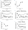
Systemic and intraplantar administration of KT109 inhibited mechanical hyperalgesia in HbSS mice. (A) HbSS mice exhibited significant mechanical hyperalgesia prior to drug injections (BL, baseline). KT109 (30 mg) was administered by intraperitoneal injection. Vehicle was DMSO:Tween 80:saline (30:1:69 v:v:v). A reduction in hyperalgesia occurred at 60 min and persisted through the 3 h testing period (F[12,90]=3.72, P<0.001 for treatment, n=4-7 mice/group, 2-way repeated measures analysis of variance [ANOVA]). KT109 had no effect in HbAA mice, and the vehicle was without effect in either strain (P=1.0 in HbAA mice, P=0.57 in HbSS mice, 2-way repeated measures ANOVA). *Different from vehicle in HbAA mice at P<0.001, #Different from vehicle in HbAA mice at P<0.05, different from vehicle in HbSS mice at P<0.001; +different from vehicle in HbSS at P<0.05 (2-way repeated measures ANOVA with Bonferroni t test). (B) A dose-dependent effect was observed for 3-100 mg KT109 (F[5, 33]=5.341, P<0.001 for treatment, n=4-8 mice/dose, one-way ANOVA). Data for doses were converted to a percent of the maximum possible effect (%MPE). Percent MPE was defined as the average response in the vehicle-treated HbSS mice (V HbSS) minus the post-drug (PD) response in the KT109-treated HbSS mice divided by the average response in the vehicle-treated HbSS mice (V HbSS) minus the average response in vehicle-treated HbAA (V HbAA) mice and multiplied by 100%: %MPE = (V HbSS – PD HbSS)/(V HbSS – V HbAA) x 100%. A dose response analysis confirmed that the dose of 30 mg (intraperitoneal) was the minimally effective dose. The EC50 was 13.1 mg (95% confidence interval: 0.61-283 mg) (GraphPad Prism). Doses were plotted on a log scale. (C) Vehicle was DMSO:Tween 80:saline (13:0.5:86.5 v:v:v). HbSS mice injected with vehicle remained different from HbAA mice injected with vehicle throughout the testing period. KT109 (3 mg, intraplantar) blocked mechanical hyperalgesia ipsilateral to the injection through 24 h (F[3,168]=30.4, P<0.001 for treatment effect, n=5-8 mice/group, 2-way repeated measures ANOVA). *Different from HbSS mice injected with KT109 at P<0.05, **different at P<0.001, different from HbAA mice at P<0.001 (2-way repeated measures ANOVA with Bonferroni t test). (D) Mechanical hyperalgesia was also blocked in the paw contralateral to the injection (F[1,88]=83.2, P<0.001 for treatment effect, 2-way repeated measures ANOVA), but the effect was not observed until 90 min after intraplantar injection of the drug (#different from vehicle in HbSS mice at P<0.001). Limited data from (A) are included for perspective. (E) Testing doses of 1, 3 and 10 mg (intraplantar) confirmed that 3 mg was the minimum effective dose to reduce mechanical hyperalgesia ipsilateral to the injection in HbSS mice. *Different from vehicle at P<0.001, one-way ANOVA with Bonferroni t test; n=5-8 mice/dose. Doses were plotted on a log scale. (F) The analog KT195 did not reduce mechanical sensitivity ipsilateral to the injection in HbSS mice when administered at the effective dose of KT109 (3 mg, intraplantar). (F[12,100]=4.2, P<0.001 for treatment effect, n=6-8 mice/group, 2-way repeated measures ANOVA). *Different from KT195 and vehicle at P<0.001, two-way ANOVA repeated measures with Bonferroni t test. BL: baseline; i.p.: intraperitoneal; i.pl.: intraplantar.
Figure 6.
Figure 6.
Intraplantar administration of KT109 reduced heat hyperalgesia in HbSS mice. (A) HbSS mice had a shorter latency to withdraw from the heat stimulus prior to drug injection. *Different from the same treatment group in HbAA mice at P<0.001 (F[3,60]=20.7, n=4-6 mice/group, 2-way repeated measures analysis of variance [ANOVA] with Bonferroni t test). HbSS mice injected with KT109 maintained this difference from HbAA mice injected with KT109 60 min after drug administration in the paw ipsilateral to the injection, but heat hyperalgesia in HbSS mice was blocked at 2 and 3 h after drug administration (different from HbSS mice treated with vehicle at P<0.05). (B) Heat hyperalgesia was also blocked in the contralateral hind paw following intraplantar injection of KT109 into the opposite hind paw in a parallel time course (different from HbSS mice treated with vehicle at P<0.05). Limited data from (A) are included for perspective. (C) Testing doses of 1, 3 and 10 mg (intraplantar) confirmed that 3 mg was the minimum effective dose to reduce thermal hyperalgesia ipsilateral to the injection in HbSS mice. *Different from vehicle at P<0.05, one-way ANOVA with Bonferroni t test; n=6-8 mice/dose. Doses are plotted on a log scale.
Figure 7.
Figure 7.
Biochemical pathways involved in the modulation of pain by KT109 in sickle cell disease. In contrast to HbAA mice, hyperalgesic HbSS mice demonstrated an increase in diacylglycerol lipase β (DAGLβ) in blood cells and 2-arachidonoylglycerol (2-AG) in plasma. High levels of cyclooxygenase-2 (COX-2) oxidize 2-AG to generate the pro-nociceptive lipid mediator prostaglandin E2-glyceryl ester (PGE2-G), which causes pain by sensitizing nociceptors. By inhibiting the enzyme activity of DAGLβ, KT109 reduces the accumulation of 2-AG, a target for COX-2, and thus blocks hyperalgesia in HbSS mice. DAG: diacylglycerol.

Comment in

References

    1. Ballas SK, Gupta K, Adams-Graves P. Sickle cell pain: a critical reappraisal. Blood. 2012;120(18):3647-3656. - PubMed
    1. Brandow AM, Stucky CL, Hillery CA, Hoffmann RG, Panepinto JA. Patients with sickle cell disease have increased sensitivity to cold and heat. Am J Hematol. 2013;88(1):37-43. - PMC - PubMed
    1. Lutz B, Meiler SE, Bekker A, Tao YX. Updated mechanisms of sickle cell disease-associated chronic pain. Transl Perioper Pain Med. 2015;2(2):8-17. - PMC - PubMed
    1. Brandow AM, Zappia KJ, Stucky CL. Sickle cell disease: a natural model of acute and chronic pain. Pain. 2017;158(Suppl 1):S79-S84. - PMC - PubMed
    1. Gupta K, Jahagirdar O, Gupta K. Targeting pain at its source in sickle cell disease. Am J Physiol Regul Integr Comp Physiol. 2018;315(1):R104-R112. - PMC - PubMed

Publication types