. 2022 Jun 21;145(25):1853-1866.
doi: 10.1161/CIRCULATIONAHA.122.059863. Epub 2022 May 26.

Fine-Tuning Cardiac Insulin-Like Growth Factor 1 Receptor Signaling to Promote Health and Longevity

Affiliations

Fine-Tuning Cardiac Insulin-Like Growth Factor 1 Receptor Signaling to Promote Health and Longevity

Mahmoud Abdellatif et al. Circulation. .

Abstract

Background: The insulin-like growth factor 1 (IGF1) pathway is a key regulator of cellular metabolism and aging. Although its inhibition promotes longevity across species, the effect of attenuated IGF1 signaling on cardiac aging remains controversial.

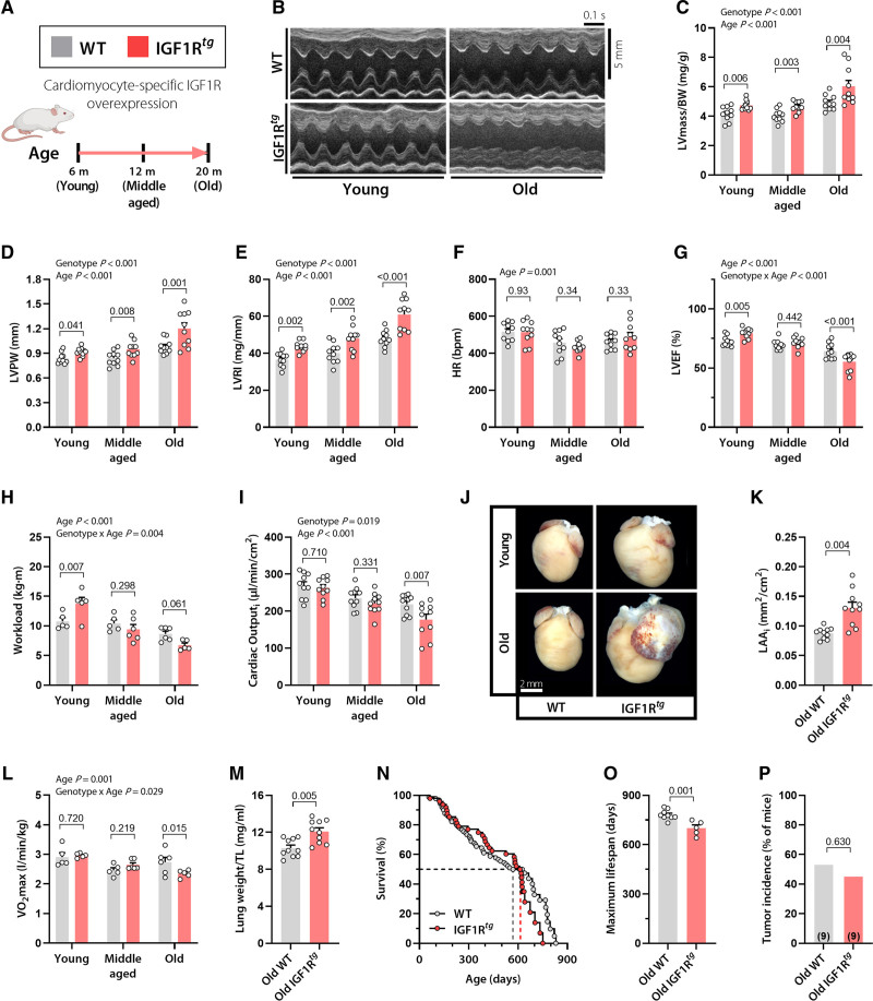
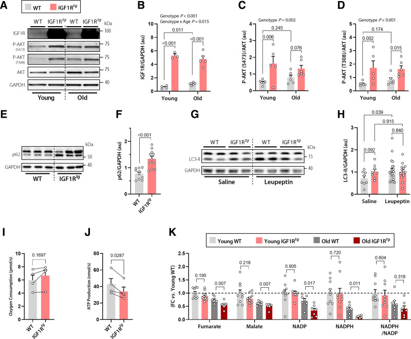
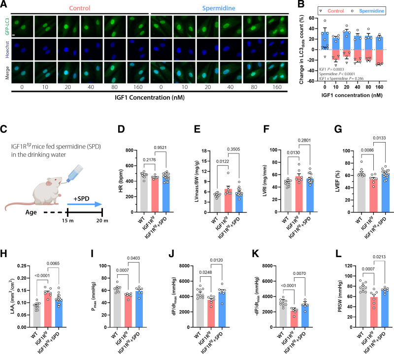
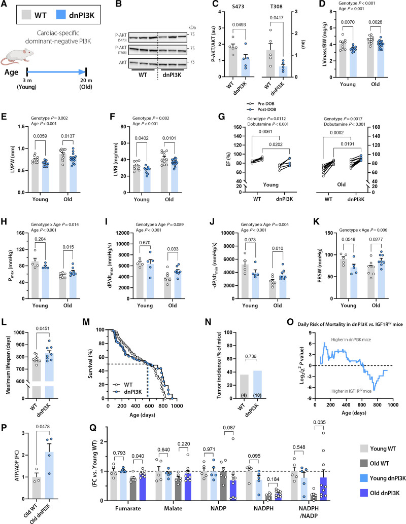
Methods: We performed a lifelong study to assess cardiac health and lifespan in 2 cardiomyocyte-specific transgenic mouse models with enhanced versus reduced IGF1 receptor (IGF1R) signaling. Male mice with human IGF1R overexpression or dominant negative phosphoinositide 3-kinase mutation were examined at different life stages by echocardiography, invasive hemodynamics, and treadmill coupled to indirect calorimetry. In vitro assays included cardiac histology, mitochondrial respiration, ATP synthesis, autophagic flux, and targeted metabolome profiling, and immunoblots of key IGF1R downstream targets in mouse and human explanted failing and nonfailing hearts, as well.

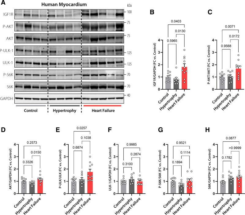
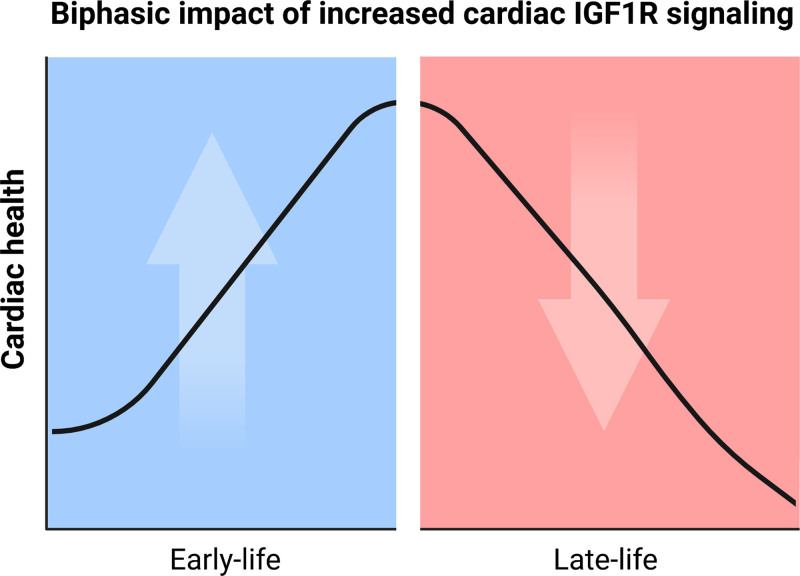
Results: Young mice with increased IGF1R signaling exhibited superior cardiac function that progressively declined with aging in an accelerated fashion compared with wild-type animals, resulting in heart failure and a reduced lifespan. In contrast, mice with low cardiac IGF1R signaling exhibited inferior cardiac function early in life, but superior cardiac performance during aging, and increased maximum lifespan, as well. Mechanistically, the late-life detrimental effects of IGF1R activation correlated with suppressed autophagic flux and impaired oxidative phosphorylation in the heart. Low IGF1R activity consistently improved myocardial bioenergetics and function of the aging heart in an autophagy-dependent manner. In humans, failing hearts, but not those with compensated hypertrophy, displayed exaggerated IGF1R expression and signaling activity.

Conclusions: Our findings indicate that the relationship between IGF1R signaling and cardiac health is not linear, but rather biphasic. Hence, pharmacological inhibitors of the IGF1 pathway, albeit unsuitable for young individuals, might be worth considering in older adults.

Keywords: aging; autophagy; cardiomyopathies; insulin-like growth factor 1; mitochondria; mouse; phosphatidylinositol 3-kinases.


Figures

Figure 1.
Figure 1.
Cardiac overexpression of IGF1R improves ejection fraction and effort tolerance in early adulthood, but causes heart failure and premature mortality late in life. A, Male mice with cardiomyocyte-specific overexpression of the human IGF1 receptor (IGF1Rtg) and their wild-type (WT) littermates (FVB/N background) were subjected to a comprehensive assessment of cardiac function and structure at the indicated age. B, Representative echocardiography-derived left ventricular (LV) M-mode tracings; C, LV mass normalized to body weight (BW); D, LV posterior wall thickness (LVPW); E, LV remodeling index (LVRI); F, heart rate (HR); and G, LV ejection fraction (LVEF) in 6-, 12-, and 20-month-old IGF1Rtg and WT mice (n=10 mice per group). H, Maximum workload during exercise tolerance testing in 3-, 12-, and 20-month-old IGF1Rtg and WT mice (n=5–6 mice per group). I, Body surface area–normalized cardiac output (cardiac index), derived by echocardiography, in IGF1Rtg and WT mice at the age of 6, 12, and 20 months (n=10 mice per group). J and K, Representative heart photomicrographs (J) and body surface area–normalized left atrial area (LAAi; K) in 20-month-old IGF1Rtg and WT mice (n=10 mice per group). L, Maximum oxygen consumption (Vo2max) during exercise tolerance testing in IGF1Rtg and WT mice at the age of 3, 12, and 20 months (n=5–6 mice per group). M, Lung weight normalized to tibia length (TL) in 20-month-old IGF1Rtg and WT mice (n=10 mice per group). N, Kaplan-Meier survival analysis of IGF1Rtg and WT mice (n=48/90 mice per group, respectively). Median survival is depicted by the dashed lines (please see also Table S3 for details). O, Maximum lifespan calculated as the average lifespan of the longest-lived decile in IGF1Rtg and WT mice (n=5/9 mice per group, respectively). P, Tumor incidence detected by gross necropsy in IGF1Rtg and WT mice (20/17 mice per group, respectively). Number in brackets indicates the number of mice that developed tumors. Indicated P values on top of panels (C–I and L) represent factor comparisons by 2-way ANOVA including genotype and age as fixed factors, followed by simple main effects analysis of pairwise comparisons between IGF1Rtg and their age-matched WT controls. Other P values were calculated by the Welch t test (K, M, and O) or χ2 test (P). Bars and error bars show means and SEM, respectively, with individual data points superimposed. IGF1 indicates insulin-like growth factor 1; IGF1R, IGF1 receptor; and IGF1Rtg mice, mice overexpressing human IGF1R specifically in cardiomyocytes.
Figure 2.
Figure 2.
Impaired autophagy and mitochondrial oxidative capacity underlie the detrimental effect of cardiac IGF1R signaling in aged IGF1Rtg mice. A through D, Representative Western blots (A) and quantification (B) of cardiac IGF1 receptor (IGF1R) expression normalized to GAPDH, and AKT phosphorylation at Ser473 (C) and Thr308 normalized to total AKT expression (D) in 6-month-old (young) and 20-month-old (old) IGF1Rtg and WT mice (n=3–6 mice per group). E and F, Representative Western blots (E) and quantification of (F) the autophagy substrate p62 normalized to GAPDH in cardiac lysates of 12-month-old IGF1Rtg and WT mice (n=11/10 mice per group, respectively). G and H, Representative Western blots (G) and quantification of (H) the lipidated form of the autophagy marker LC3-II in the hearts of 12-month-old IGF1Rtg and WT mice that were treated with the protease inhibitor leupeptin (n=14/21 mice per group, respectively) or saline (n=8 mice per group). I and J, Paired assessment of oxygen consumption rate (I) and corresponding ATP production (J) in cardiac mitochondria isolated from 20-month-old IGF1Rtg and WT mice (n=4 mice per group). K, Relative difference in the tricarboxylic acid cycle intermediates, fumarate and malate, and the ratio between reduced and oxidized forms of the cofactor nicotinamide adenine dinucleotide phosphate (NADPH/NADP) in young (3 months old) and old (20 months old) IGF1Rtg and WT mice (n=7–10 mice per group). Indicated P values on top of panels (B–D and H) represent factor comparisons by 2-way ANOVA including genotype and age (B–D) or genotype and treatment (H) as fixed factors; the following simple main effects denote pairwise comparisons between IGF1Rtg and their respective WT controls within a specific age or treatment. Other P values were calculated by unpaired Welch t test or Mann-Whitney test (F and K) or paired Student t test (I and J). Bars and error bars show means and SEM, respectively, with individual data points superimposed. FC indicates fold change; IGF1, insulin-like growth factor 1; IGF1R, IGF1 receptor; IGF1Rtg mice, mice overexpressing human IGF1R specifically in cardiomyocytes; and WT, wild-type.
Figure 3.
Figure 3.
The autophagy inducer spermidine ameliorates the cardiac phenotype of aged IGF1Rtg mice. A and B, Representative confocal images (A) and quantification (B) of autophagic activity assessed by GFP-LC3 positive dots in GFP-LC3–expressing H9c2 cells treated with increasing concentrations of IGF1 in the presence or absence of spermidine (6.25 µmol/L) for 6 hours. Scale bar, 10 µm (n=4 biological replicates per condition). C, Schematic representation of spermidine (SPD) feeding protocol to male IGF1Rtg mice. SPD (3 mmol/L) was added to the drinking water starting at the age of 15 months, and after 5 months cardiac parameters were assessed. D through H, Echocardiography-derived (D) heart rate (HR), LV mass normalized to body weight (BW; E), LV remodeling index (LVRI; F), LV ejection fraction (LVEF; G), and body surface area–normalized left atrial area (LAAi; H) in 20-month-old spermidine-treated and control IGF1Rtg and WT mice (n=15/7/11 mice per group, respectively). I through L, Invasively measured left ventricular maximum pressure (Pmax; I), maximum rate of pressure rise (dP/dtmax; J), maximum rate of pressure decay (–dP/dtmin; K), and preload recruitable stroke work (PRSW; L) in 20-month-old spermidine-treated and control IGF1Rtg and WT mice (n=7/7/10 mice per group, respectively). Indicated P values on top of B represent factor comparisons by 2-way ANOVA including IGF1 concentration and spermidine treatment as fixed factors. Other P values were calculated by ANOVA with the Dunnett post hoc test. Bars and error bars show means and SEM, respectively, with individual data points superimposed. GFP-LC3 indicates Green Fluorescent Protein-Microtubule-associated Protein 1A/1B-Light Chain 3; IGF1, insulin-like growth factor 1; IGF1R, IGF1 receptor; IGF1Rtg mice, mice overexpressing human IGF1R specifically in cardiomyocytes; LV, left ventricular; and WT, wild-type.
Figure 4.
Figure 4.
Reduced cardiomyocyte IGF1R signaling in dnPI3K mice delays cardiac growth, but protects from age-related decline in cardiac function. A, Dominant negative PI3K (dnPI3K) male mice with reduced cardiomyocyte-specific IGF1R signaling and their WT littermates (FVB/N background) were subjected to a comprehensive assessment of cardiac function and structure at the age of 3 months (young) and 20 months (old). B and C, Representative Western blots (B) and quantification of (C) cardiac AKT phosphorylation at Ser473 and Thr308 normalized to total AKT expression in 10-month-old dnPI3K and WT mice (n=5 mice per group). D through F, Echocardiographic assessment of left ventricular (LV) mass normalized to body weight (BW; D), LV posterior wall thickness (LVPW; E), and LV remodeling index (LVRI; F) in young and old dnPI3K and WT mice (n=10 young and 12–15 old mice per genotype). G, LV ejection fraction (LVEF) before and after β-adrenergic stimulation by intraperitoneal injection of dobutamine (1.5 mg/kg IP) in young (Left) and old (Right) dnPI3K and WT mice (n=5 young and n=10 old mice per genotype). H through K, Invasive assessment of LV maximum pressure (Pmax; H), maximum rate of pressure rise (dP/dtmax; I), maximum rate of pressure decay (dP/dtmin; J), and preload recruitable stroke work (PRSW; K) in young and old dnPI3K and WT mice (n=5–8 mice per group). L, Maximum lifespan was calculated as the average lifespan of the longest-lived decile in dnPI3K and WT mice (n=8/9 mice per genotype, respectively). M, Kaplan-Meier survival analysis in dnPI3K and WT mice (n=80/90 mice per genotype, respectively). The same WT control group as in Figure 1N was used. Median survival is indicated by the corresponding dashed lines (also see Table S3 for further details). N, Tumor incidence detected by gross necropsy in dnPI3K and WT mice (26/11 mice per genotype, respectively). Number in brackets indicates the number of mice that developed tumors. O, Daily mortality risk in dnPI3K compared with IGF1Rtg mice (n=80/48 mice per genotype, respectively). P, Cardiac abundance of ATP/ADP ratio in 20-month-old dnPI3K and WT mice (n=4–5 mice per genotype). Q, Relative difference in the tricarboxylic acid cycle intermediates, fumarate and malate, and the ratio between reduced and oxidized forms of the cofactor nicotinamide adenine dinucleotide phosphate (NADPH/NADP) in young (3 months old) and old (20 months old) dnPI3K and WT mice (n=5–9 mice per group). Indicated P values on top of D through K represent factor comparisons by 2-way ANOVA including genotype and age as fixed factors (D–F, H–K) or genotype and dobutamine (G), followed by simple main effects analysis of pairwise comparisons between dnPI3K and their age- or treatment-matched WT controls. Other P values were calculated by the Welch t test or Mann-Whitney test, as appropriate (L, P through Q) or χ2 test (N). Bars and error bars show means and SEM, respectively, with individual data points superimposed. dnPI3K indicates mice harboring an inactivated (dominant negative, dn) p110α isoform of phosphoinositide 3-kinase; IGF1, insulin-like growth factor 1; IGF1R, IGF1 receptor; IGF1Rtg mice, mice overexpressing human IGF1R specifically in cardiomyocytes; and WT, wild-type.
Figure 5.
Figure 5.
Increased IGF1R signaling in human failing hearts. Representative Western blots (A) and immunoblot analysis of cardiac IGF1 receptor (IGF1R) expression (B), AKT phosphorylation at Thr308 normalized to total AKT expression (C), AKT expression (D), ULK-1 phosphorylation normalized to total ULK-1 expression (E), ULK-1 expression (F), S6K phosphorylation normalized to total S6K expression (G), and S6K expression (H) in left ventricular samples obtained from failing and nonfailing human hearts with or without echocardiographic evidence of hypertrophy (n=9/10/10 hearts in Control, Hypertrophy, and Heart Failure, respectively). GAPDH was used as a loading control. Indicated P values were calculated by Welch test with Dunnett T3 post hoc (B and E), ANOVA with Tukey post hoc (C, D, F, G) or Kruskal-Wallis-test with Dunn post hoc (H). Bars and error bars show means and SEM, respectively, with individual data points superimposed. FC indicates fold change; IGF1, insulin-like growth factor 1; IGF1R, IGF1 receptor; and IGF1Rtg mice, mice overexpressing human IGF1R specifically in cardiomyocytes.
Figure 6.
Figure 6.
Graphical summary of the role of cardiomyocyte IGF1R signaling in regulating cardiac health during the course of life. IGF1R signaling proved crucial for cardiac homeostasis during early life as young IGF1Rtg mice exhibited cardiac benefits in association with increased IGF1R signaling, whereas IGF1R activity was deleterious for cardiac function in aged IGF1Rtg mice. Conversely, reduced cardiac IGF1R signaling in dnPI3K mice was associated with lower cardiac performance and increased risk of mortality in early life, but extended cardiac healthspan and maximum longevity later in life. dnPI3K indicates mice harboring an inactivated (dominant negative, dn) p110α isoform of phosphoinositide 3-kinase; IGF1, insulin-like growth factor 1; IGF1R, IGF1 receptor; and IGF1Rtg mice, mice overexpressing human IGF1R specifically in cardiomyocytes.

Comment in

References

    1. Johnson SC. Nutrient sensing, signaling and ageing: the role of IGF-1 and mTOR in ageing and age-related disease. Subcell Biochem. 2018;90:49–97. doi: 10.1007/978-981-13-2835-0_3 - PubMed
    1. Milman S, Huffman DM, Barzilai N. The somatotropic axis in human aging: framework for the current state of knowledge and future research. Cell Metab. 2016;23:980–989. doi: 10.1016/j.cmet.2016.05.014 - PMC - PubMed
    1. Mao K, Quipildor GF, Tabrizian T, Novaj A, Guan F, Walters RO, Delahaye F, Hubbard GB, Ikeno Y, Ejima K, et al. . Late-life targeting of the IGF-1 receptor improves healthspan and lifespan in female mice. Nat Commun. 2018;9:2394. doi: 10.1038/s41467-018-04805-5 - PMC - PubMed
    1. Bokov AF, Garg N, Ikeno Y, Thakur S, Musi N, DeFronzo RA, Zhang N, Erickson RC, Gelfond J, Hubbard GB, et al. . Does reduced IGF-1R signaling in Igf1r+/– mice alter aging? PLoS One. 2011;6:e26891. doi: 10.1371/journal.pone.0026891 - PMC - PubMed
    1. Xu J, Gontier G, Chaker Z, Lacube P, Dupont J, Holzenberger M. Longevity effect of IGF-1R+/– mutation depends on genetic background-specific receptor activation. Aging Cell. 2014;13:19–28. doi: 10.1111/acel.12145 - PMC - PubMed

Publication types

Substances