. 2022 Jul;298(7):102066.
doi: 10.1016/j.jbc.2022.102066. Epub 2022 May 23.

Maintaining the thyroid gland in mutant thyroglobulin-induced hypothyroidism requires thyroid cell proliferation that must continue in adulthood

Affiliations

Maintaining the thyroid gland in mutant thyroglobulin-induced hypothyroidism requires thyroid cell proliferation that must continue in adulthood

Xiaohan Zhang et al. J Biol Chem. 2022 Jul.

Abstract

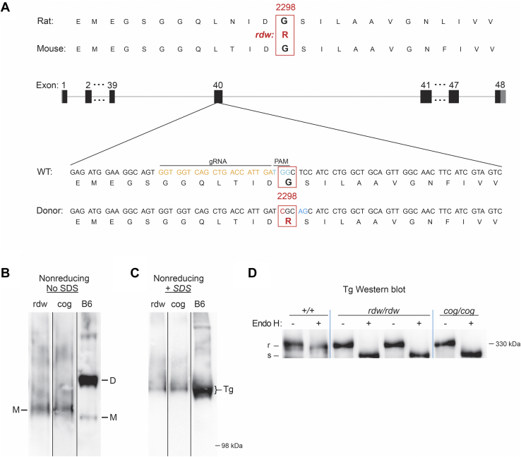
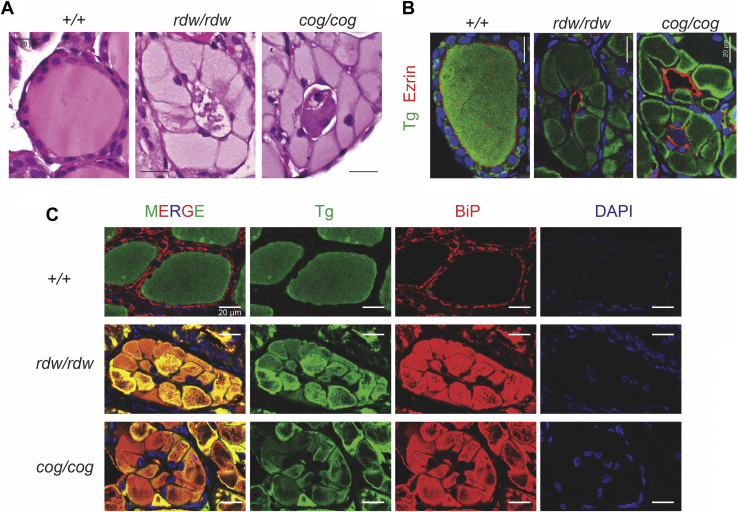
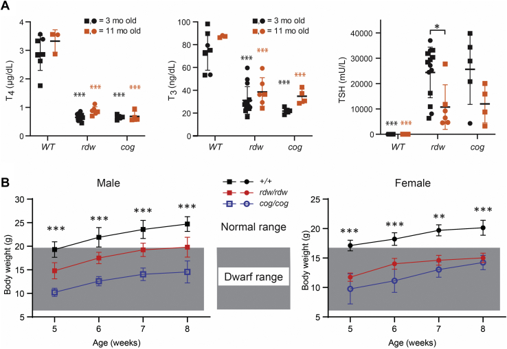
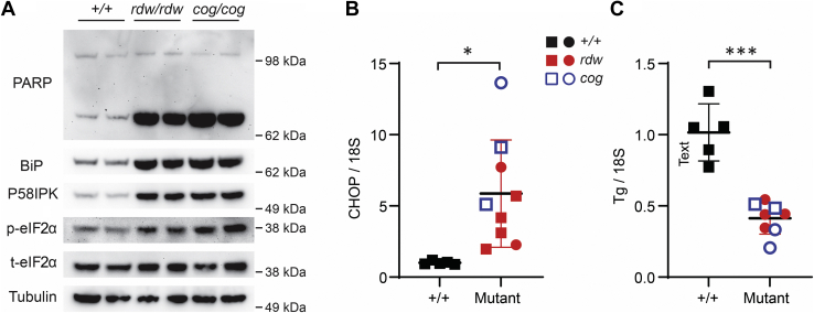
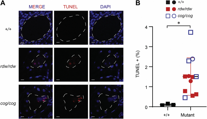
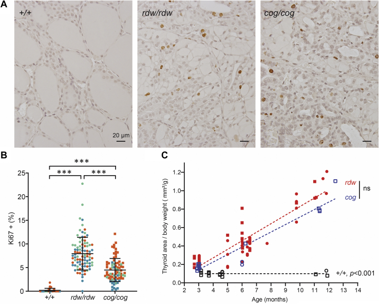
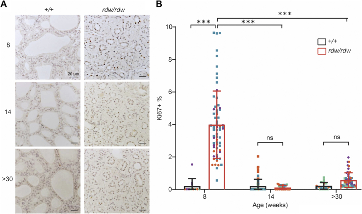
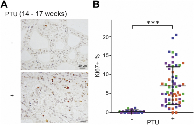
Congenital hypothyroidism with biallelic thyroglobulin (Tg protein, encoded by the TG gene) mutation is an endoplasmic reticulum (ER) storage disease. Many patients (and animal models) grow an enlarged thyroid (goiter), yet some do not. In adulthood, hypothyroid TGcog/cog mice (bearing a Tg-L2263P mutation) exhibit a large goiter, whereas adult WIC rats bearing the TGrdw/rdw mutation (Tg-G2298R) exhibit a hypoplastic thyroid. Homozygous TG mutation has been linked to thyroid cell death, and cytotoxicity of the Tg-G2298R protein was previously thought to explain the lack of goiter in WIC-TGrdw/rdw rats. However, recent studies revealed that TGcog/cog mice also exhibit widespread ER stress-mediated thyrocyte death, yet under continuous feedback stimulation, thyroid cells proliferate in excess of their demise. Here, to examine the relative proteotoxicity of the Tg-G2298R protein, we have used CRISPR-CRISPR-associated protein 9 technology to generate homozygous TGrdw/rdw knock-in mice in a strain background identical to that of TGcog/cog mice. TGrdw/rdw mice exhibit similar phenotypes of defective Tg protein folding, thyroid histological abnormalities, hypothyroidism, and growth retardation. TGrdw/rdw mice do not show evidence of greater ER stress response or stress-mediated cell death than TGcog/cog mice, and both mouse models exhibit sustained thyrocyte proliferation, with comparable goiter growth. In contrast, in WIC-TGrdw/rdw rats, as a function of aging, the thyrocyte proliferation rate declines precipitously. We conclude that the mutant Tg-G2298R protein is not intrinsically more proteotoxic than Tg-L2263P; rather, aging-dependent difference in maintenance of cell proliferation is the limiting factor, which accounts for the absence of goiter in adult WIC-TGrdw/rdw rats.

Keywords: ER stress; aging; cell death; protein misfolding; secretory pathway.


Conflict of interest statement

Conflict of interest The authors declare that they have no conflicts of interest with the contents of this article.

Figures

Figure 1
Figure 1
Generation of TGrdw/rdwmice.A, schematic illustration of CRISPR/Cas9-mediated gene editing to knock-in rdw (Tg-G2298R) mutation within the exon 40 of the mouse TG gene. The target Tg protein sequences and the target site of gRNA are indicated. B and C, polyacrylamide gel electrophoresis of thyroid homogenates from B6, TGrdw/rdw, and TGcog/cog mice in the absence (B) or the presence (C) of SDS, followed by immunoblotting with an mAb anti-Tg (three repeats). D, endoglycosidase H digest and Tg Western blotting of thyroid from B6, TGrdw/rdw, and TGcog/cog mice (n = 3 animals per group). Cas9, CRISPR-associated protein 9; gRNA, guide RNA; mAb, monoclonal antibody; TG, thyroglobulin gene; Tg, thyroglobulin protein.
Figure 2
Figure 2
Thyroid histology of TGrdw/rdwmice.A, representative H&E of thyroid glands from B6, TGrdw/rdw, and TGcog/cog mice (n = 5–8 animals per group), showing thyrocyte distention in TGrdw/rdw and TGcog/cog mice. The scale bars represent 20 μm. B, immunofluorescence of anti-Tg (green) and antiezrin (red) of thyroid glands from B6, TGrdw/rdw, and TGcog/cog mice (n = 5 animals per group), with DAPI counterstaining (blue). The scale bars represent 20 μm. C, immunofluorescence of Tg (green) and anti-BiP (red) of thyroid glands from B6, TGrdw/rdw, and TGcog/cog mice (n = 5 animals per group), with DAPI counterstaining (blue). The scale bars represent 20 μm. BiP, immunoglobulin heavy-chain binding protein; DAPI, 4′,6-diamidino-2-phenylindole; TG, thyroglobulin gene; Tg, thyroglobulin protein.
Figure 3
Figure 3
Hypothyroidism of TGrdw/rdwmice.A, serum total T4, T3, and TSH levels of B6, TGrdw/rdw, and TGcog/cog mice at 3 months (black) and 11 months (red) of age (males shown as squares and females as circles). Data are mean ± SD; ∗p < 0.05, #p < 0.001 compared with B6 mice of 3 months old, &p < 0.001 compared with B6 mice of 11 months old. B, body weight of B6 (black), TGrdw/rdw, (red) and TGcog/cog mice (blue). The shaded area indicates the body weights of dwarf mice. Data are mean ± SD; ∗∗p < 0.01, ∗∗∗p < 0.001 compared with B6 mice. T3, triiodothyronine; T4, thyroxine; TG, thyroglobulin gene; TSH, thyroid stimulating hormone.
Figure 4
Figure 4
Chronic ER stress in the thyroid glands of TGrdw/rdwmice.A, BiP, p58ipk, phospho-eIF2α, and PARP (by Western blotting) in the thyroid glands of B6, TGrdw/rdw, and TGcog/cog mice (n = 3 animals per group, two of which are shown in the panel). B, CHOP mRNA levels (normalized to 18S) in the thyroid glands of B6, TGrdw/rdw (purple), and TGcog/cog (orange) mice (n = 3–6 animals per group; each point represents a single animal; square = male, circle = female). Data are shown as mean ± SD; ∗p < 0.05. C, Tg mRNA levels (normalized to 18S) in the thyroid glands of B6 (black symbols), TGrdw/rdw (red symbols), and TGcog/cog (blue symbols) mice (n = 4–5 animals per group; each point represents a single animal; square = male, circle = female). Data are shown as mean ± SD; ∗∗∗p < 0.001. BiP, immunoglobulin heavy-chain binding protein; CHOP, CCAAT/enhancer-binding protein homologous protein; eIF2α, eukaryotic initiation factor 2α; ER, endoplasmic reticulum; PARP, poly(ADP-ribose) polymerase; TG, thyroglobulin gene; Tg, thyroglobulin protein.
Figure 5
Figure 5
Thyroid cell death in TGrdw/rdwmice.A, representative TUNEL labeling (red) with DAPI counterstain (blue) in the thyroid sections of B6, TGrdw/rdw, and TGcog/cog mice (n = 3–7 animals per group). A dashed white line delimits the thyroid follicle lumen. The scale bars represent 10 μm. B, quantification of A presented as TUNEL-positive nuclei in the proportion of total nuclei in the thyroids of B6 (black symbols), TGrdw/rdw (red symbols), and TGcog/cog (blue symbols) mice (n = 3–7 animals per group; each point represents a single animal; square = male, circle = female). Data are mean ± SD; ∗p < 0.05. DAPI, 4′,6-diamidino-2-phenylindole; TG, thyroglobulin gene.
Figure 6
Figure 6
Thyroid cell proliferation in TGrdw/rdwmice.A, Ki67 immunohistochemistry of the thyroid gland of WT, TGrdw/rdw, and TGcog/cog mice (11 months). There are sustained thyroid cell proliferation in both TGrdw/rdw and TGcog/cog mice. The scale bars represent 20 μm. B, quantification of images like those shown in A, presented as Ki67-positive nuclei as a proportion of total nuclei in thyroid images from 11-month-old WT, TGrdw/rdw, and TGcog/cog mice (n = 3 animals per group; each color represents a single animal; each point is an independent section; square = male, circle = female). Data are mean ± SD; ∗∗∗p < 0.001. C, thyroid gland size (normalized to body weight) as a function of age (square = male, circle = female). A dashed line shows the linear regression of the thyroid size in WT, TGrdw/rdw, and TGcog/cog mice. No significance difference was observed between thyroid glands of TGrdw/rdw and TGcog/cog mice. TG, thyroglobulin gene.
Figure 7
Figure 7
Thyroid cell proliferation in WIC-TGrdw/rdwrats.A, Ki67 immunohistochemistry of the thyroid gland of WT and WIC-TGrdw/rdw rats at 8 weeks, 14 weeks, and over 30 weeks of age. The scale bars represent 20 μm. B, quantification of immunostained sections like those shown in A, presented as Ki67-positive nuclei in the proportion of total nuclei in the thyroids of WT WIC and WIC-TGrdw/rdw rats (n = 3–4 animals per group; each color represents a single animal; each point is an independent section; square = male, circle = female). Data are mean ± SD; ∗∗∗p < 0.001. TG, thyroglobulin gene.
Figure 8
Figure 8
Thyroid cell proliferation in PTU-induced hypothyroid adult WT rats.A, Ki67 immunohistochemistry of the thyroid gland of control WT and PTU-treated WT rats at 17 weeks of age. Treated rats received PTU chow at 14 weeks of age for 21 days. The scale bars represent 20 μm. B, quantification of A presented as Ki67-positive nuclei in the proportion of total nuclei in the thyroids of control WT and PTU-treated WT rats (n = 3–4 animals per group; each color represents a single animal; each point is an independent section, males shown as squares and females as circles). Data are mean ± SD; ∗∗∗p < 0.001. PTU, propylthiouracil.

Similar articles

Cited by

References

    1. Brix K., Qatato M., Szumska J., Venugopalan V., Rehders M. In: The Thyroid and its Diseases: A Comprehensive Guide for the Clinician. Luster M., Duntas L.H., Wartofsky L., editors. Springer International Publishing; Cham: 2019. “Thyroglobulin storage, processing and degradation for thyroid hormone liberation”; pp. 25–48.
    1. Holzer G., Morishita Y., Fini J.B., Lorin T., Gillet B., Hughes S., et al. Thyroglobulin represents a novel molecular architecture of vertebrates. J. Biol. Chem. 2016;291:16553–16566. - PMC - PubMed
    1. Di Jeso B., Arvan P. Thyroglobulin from molecular and cellular biology to clinical endocrinology. Endocr. Rev. 2016;37:2–36. - PMC - PubMed
    1. Carvalho D.P., Dupuy C. Thyroid hormone biosynthesis and release. Mol. Cell Endocrinol. 2017;458:6–15. - PubMed
    1. Dunn J.T., Dunn A.D. The importance of thyroglobulin structure for thyroid hormone biosynthesis. Biochimie. 1999;81:505–509. - PubMed