

Promotion of corneal angiogenesis by sensory neuron-derived calcitonin gene-related peptide
- PMID: 35618042
- PMCID: PMC9428938
- DOI: 10.1016/j.exer.2022.109125
Promotion of corneal angiogenesis by sensory neuron-derived calcitonin gene-related peptide
Abstract
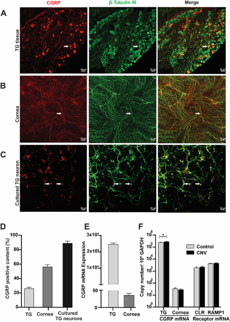
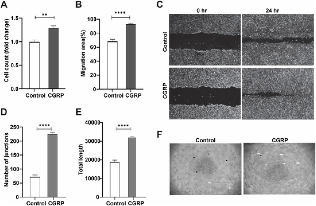
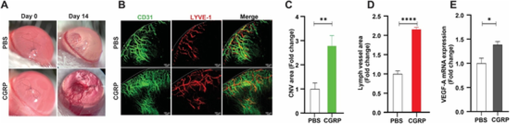
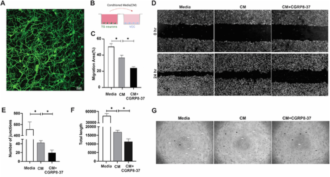
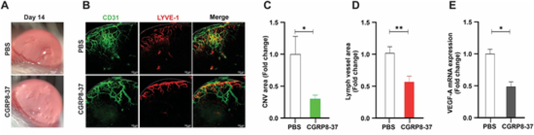
The normal cornea has no blood vessels but has abundant innervation. There is emerging evidence that sensory nerves, originated from the trigeminal ganglion (TG) neurons, play a key role in corneal angiogenesis. In the current study, we examined the role of TG sensory neuron-derived calcitonin gene-related peptide (CGRP) in promoting corneal neovascularization (CNV). We found that CGRP was expressed in the TG and cultured TG neurons. In the cornea, minimal CGRP mRNA was detected and CGRP immunohistochemical staining was exclusively co-localized with corneal nerves, suggesting corneal nerves are likely the source of CGRP in the cornea. In response to intrastromal suture placement and neovascularization in the cornea, CGRP expression was increased in the TG. In addition, we showed that CGRP was potently pro-angiogenic, leading to vascular endothelial cell (VEC) proliferation, migration, and tube formation in vitro and corneal hemangiogenesis and lymphangiogenesis in vivo. In a co-culture system of TG neurons and VEC, blocking CGRP signaling in the conditioned media of TG neurons led to decreased VEC migration and tube formation. More importantly, subconjunctival injection of a CGRP antagonist CGRP8-37 reduced suture-induced corneal hemangiogenesis and lymphangiogenesis in vivo. Taken together, our data suggest that TG sensory neuron and corneal nerve-derived CGRP promotes corneal angiogenesis.
Keywords: Calcitonin gene-related peptide; Corneal angiogenesis; Corneal innervation; Neovascularization; Neuropeptide; Trigeminal ganglion neuron.
Copyright © 2022 Elsevier Ltd. All rights reserved.
Figures
Similar articles
-
Sensory nerves promote corneal inflammation resolution via CGRP mediated transformation of macrophages to the M2 phenotype through the PI3K/AKT signaling pathway.Int Immunopharmacol. 2022 Jan;102:108426. doi: 10.1016/j.intimp.2021.108426. Epub 2021 Dec 11. Int Immunopharmacol. 2022. PMID: 34906854
-
Sensory neurons directly promote angiogenesis in response to inflammation via substance P signaling.FASEB J. 2020 May;34(5):6229-6243. doi: 10.1096/fj.201903236R. Epub 2020 Mar 12. FASEB J. 2020. PMID: 32162744 Free PMC article.
-
Sensory and sympathetic nerve sprouting in the rat cornea following neonatal administration of capsaicin.Somatosens Mot Res. 1993;10(4):377-98. doi: 10.3109/08990229309028845. Somatosens Mot Res. 1993. PMID: 7508667
-
Sensory Neuron-TRPV4 Modulates Temporomandibular Disorder Pain Via CGRP in Mice.J Pain. 2023 May;24(5):782-795. doi: 10.1016/j.jpain.2022.12.001. Epub 2022 Dec 9. J Pain. 2023. PMID: 36509176 Free PMC article. Review.
-
Current understanding of trigeminal ganglion structure and function in headache.Cephalalgia. 2019 Nov;39(13):1661-1674. doi: 10.1177/0333102418786261. Epub 2018 Jul 10. Cephalalgia. 2019. PMID: 29989427 Free PMC article. Review.
Cited by
-
Calcitonin Gene-Related Peptide Ameliorates Experimental Dry Eye Disease.Invest Ophthalmol Vis Sci. 2025 Aug 1;66(11):58. doi: 10.1167/iovs.66.11.58. Invest Ophthalmol Vis Sci. 2025. PMID: 40853308 Free PMC article.
-
Sensory Nerve Regulation via H3K27 Demethylation Revealed in Akermanite Composite Microspheres Repairing Maxillofacial Bone Defect.Adv Sci (Weinh). 2024 Aug;11(30):e2400242. doi: 10.1002/advs.202400242. Epub 2024 Jun 14. Adv Sci (Weinh). 2024. PMID: 38874525 Free PMC article.
-
Topical application of calcitonin gene-related peptide as a regenerative, antifibrotic, and immunomodulatory therapy for corneal injury.Commun Biol. 2024 Mar 4;7(1):264. doi: 10.1038/s42003-024-05934-y. Commun Biol. 2024. PMID: 38438549 Free PMC article.
-
Topical application of calcitonin gene-related peptide as a regenerative, antifibrotic, and immunomodulatory therapy for corneal injury.Res Sq [Preprint]. 2023 Aug 7:rs.3.rs-3204385. doi: 10.21203/rs.3.rs-3204385/v1. Res Sq. 2023. Update in: Commun Biol. 2024 Mar 4;7(1):264. doi: 10.1038/s42003-024-05934-y. PMID: 37609298 Free PMC article. Updated. Preprint.
-
From sensation to regulation: the diverse functions of peripheral sensory nervous system.Front Immunol. 2025 May 16;16:1575917. doi: 10.3389/fimmu.2025.1575917. eCollection 2025. Front Immunol. 2025. PMID: 40453085 Free PMC article. Review.
References
-
- Allen GS, Gross CJ, Henderson LM, Chou SN, 1976. Cerebral arterial spasm. Part 4: in vitro effects of temperature, serotonin analogues, large nonphysiological concentrations of serotonin, and extracellular calcium and magnesium on serotonin-induced contractions of the canine basilar artery. J. Neurosurg. 44 (5), 585–593. 10.3171/jns.1976.44.5.0585. - DOI - PubMed
-
- Amparo F, Sadrai Z, Jin Y, Alfonso-Bartolozzi B, Wang H, Shikari H, Ciolino JB, Chodosh J, Jurkunas U, Schaumberg DA, Dana R, 2013. Safety and efficacy of the multitargeted receptor kinase inhibitor pazopanib in the treatment of corneal neovascularization. Investig. Ophthalmol. Vis. Sci. 54 (1), 537–544. 10.1167/iovs.12-11032. - DOI - PMC - PubMed
-
- Campos X, Muñoz Y, Selman A, Yazigi R, Moyano L, Weinstein-Oppenheimer C, Lara HE, Romero C, 2007. Nerve growth factor and its high-affinity receptor trkA participate in the control of vascular endothelial growth factor expression in epithelial ovarian cancer. Gynecol. Oncol. 104 (1), 168–175. 10.1016/j.ygyno.2006.07.007. - DOI - PubMed
-
- Chiba T, Yamaguchi A, Yamatani T, Nakamura A, Morishita T, Inui T, Fukase M, Noda T, Medicine I, 1989. Calcitonin antagonist gene-related peptide receptor. Am. J. Physiol. 331–335. - PubMed
Publication types
MeSH terms
Substances
Grants and funding
LinkOut - more resources
Full Text Sources
Research Materials
Miscellaneous
