Case Reports
. 2022 May 26;22(1):262.
doi: 10.1186/s12876-022-02337-3.

A case of diverticulum of the appendiceal base resembling a submucosal tumor of the cecum under colonoscopy: a hitherto undescribed lesion

Affiliations
Case Reports

A case of diverticulum of the appendiceal base resembling a submucosal tumor of the cecum under colonoscopy: a hitherto undescribed lesion

Toshihisa Kimura et al. BMC Gastroenterol. .

Abstract

Background: Diverticulosis of the appendix is an uncommon clinical entity, and a preoperative diagnosis is often difficult. Herein we report an unusual case of appendiceal diverticulosis.

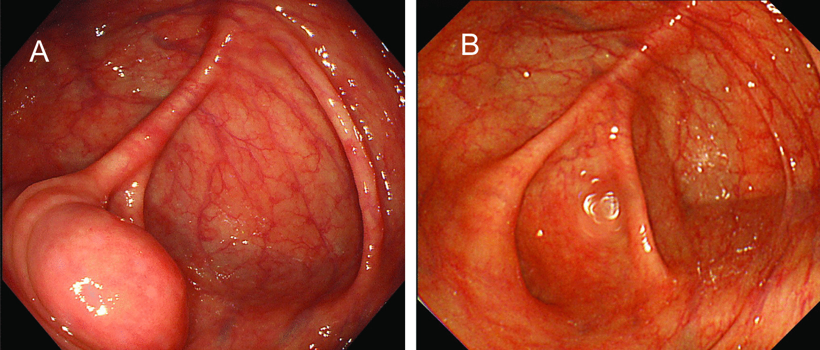
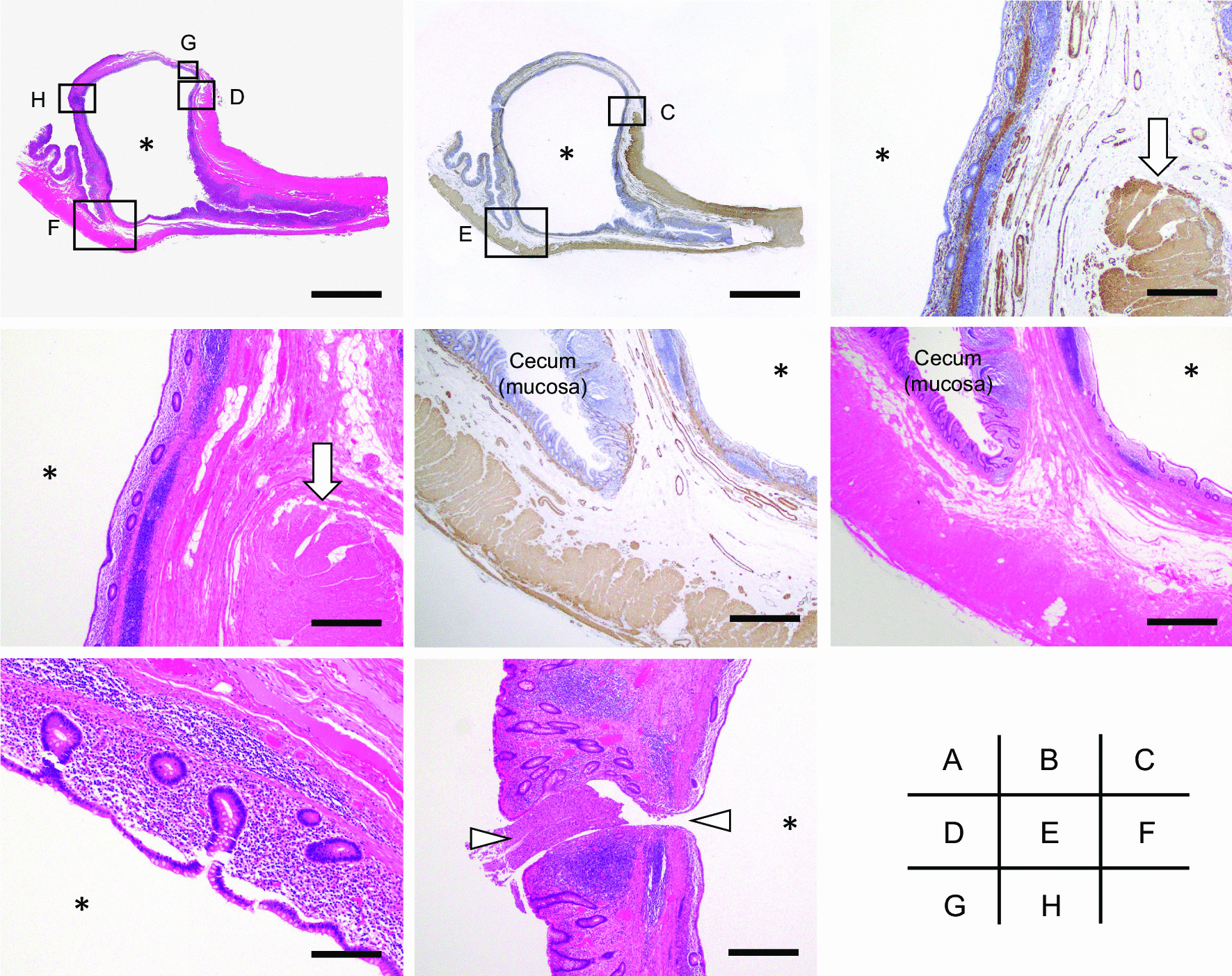
Case presentation: A 72-year-old male was referred to our hospital to examine the cause of hematochezia. A colonoscopy study showed a protruding lesion resembling a submucosal tumor (SMT), approximately 20 mm in diameter, at the site around the appendiceal orifice of the cecum. An abdominal computed tomography and magnetic resonance imaging showed a cystic lesion at the appendiceal base. The lesion was clinically diagnosed as a cystic tumor of the appendix, but the possibility of a malignant tumor could not be excluded. Therefore, a laparoscopic ileocecal resection with lymph node dissection was performed. The pathological examination of the resected specimen revealed that the lesion was a diverticulum (pseudodiverticulum) occurring solitarily at the appendiceal base, in which the mucosal layer of the appendix was invaginated into the submucosa of the adjacent cecum, thus forming an SMT-like lesion.

Conclusion: To our knowledge, this is the first case report in the English literature showing that an appendiceal diverticulum can manifest as an SMT-like lesion in the cecum. This condition should be recognized as a differential diagnosis for such lesions.

Keywords: Appendix; Case report; Colonoscopy; Diverticulosis; Diverticulum.


Conflict of interest statement

The authors declare that they have no competing interests.

Figures

Fig. 1
Fig. 1
Colonoscopy findings. Colonoscopy performed just before surgery revealed a submucosal tumor-like lesion approximately 20 mm in diameter with a smooth surface at the cecum (a). Previous colonoscopy showing a normal cecum (5 years prior to surgery, b)
Fig. 2
Fig. 2
Abdominal computed tomography (CT) scan. Enhanced abdominal CT demonstrated a cecal cystic mass (a, arrow) without significant lymph node enlargement in the abdominal cavity. The appendix was almost normal size and do not have wall thickness or an appendicolith, but had a slightly dilation of the lumen (b, arrowheads)
Fig. 3
Fig. 3
Abdominal magnetic resonance imaging (MRI) (coronal section). MRI revealed a cystic lesion at the appendiceal base with a low intensity on the T1-weighted imaging (a, arrow), and a high intensity on the T2-weighted imaging (b, arrow)
Fig. 4
Fig. 4
Macroscopic view of the resected specimen. a, b A submucosal tumor (SMT)-like lesion of the cecum (arrows). c Cut surface of the SMT-like lesion. The cut surface of the boxed area in a is shown in c. d Whole mount view of the SMT-like lesion (H&E staining). The lesion connected to the appendiceal lumen (arrowheads). The image of whole mount view was acquired by the use of a digital camera (EOS Kiss X10; Canon, Tokyo, Japan), followed by the processing using the Abode Photoshop software (Abobe, San Jose, CA). Scale bar; 1 cm (d)
Fig. 5
Fig. 5
Histological findings of the lesion. a Whole mount view of the lesion (H&E staining). b Whole mount view of the lesion (immunostaining of alpha-smooth muscle actin). The close up images of the boxed areas with alphabetical indications in a and b are shown in each corresponding figure. c, d Disruption of the muscularis propria of the appendix (arrows). e, f Photographs showing the invagination of the mucosal layer of the appendix into the submucosa of the adjacent cecum. g The lumen of the cystic lesion was covered by nonneoplastic colonic mucosa. h Communication between the lumen of the cyst and the cecum (arrowheads). Asterisks indicate the lumen of the cystic lesion. Microscopic images were acquired by the use of a light microscopy (BX43; OLYMPUS, Tokyo, Japan) equipped with the image processing software cellSens (OLYMPUS). Scale bars; 1 cm (a, b); 2 mm (c, d, h), 1 mm (e, f); 0.5 mm (g)

References

    1. Jaboury IA. Diverticulitis of the vermiform appendix. ANZ J Surg. 2007;77:803–804. doi: 10.1111/j.1445-2197.2007.04240.x. - DOI - PubMed
    1. Simpson J, Lobo DN, Spiller RC, Scholefield JH. Diverticular abscess of the appendix: report of a case and review of the literature. Dis Colon Rectum. 2003;43:832–834. doi: 10.1007/s10350-004-6664-4. - DOI - PubMed
    1. Altieri ML, Piozzi GN, Salvatori P, Mirra M, Piccolo G, Olivari N. Appendiceal diverticulitis, a rare relevant pathology: presentation of a case report and review of the literature. Int J Surg Case Rep. 2017;33:31–34. doi: 10.1016/j.ijscr.2017.02.027. - DOI - PMC - PubMed
    1. Lamps LW, Gray GF, Jr, Dilday BR, Washington MK. The coexistence of low-grade mucinous neoplasms of the appendix and appendiceal diverticula: a possible role in the pathogenesis of pseudomyxoma peritonei. Mod Pathol. 2000;13:495–501. doi: 10.1038/modpathol.3880086. - DOI - PubMed
    1. Place RJ, Simmang CL, Huber PJ. Appendiceal diverticulitis. South Med J. 2000;93:76–79. doi: 10.1097/00007611-200093010-00018. - DOI - PubMed

Publication types