. 2022 Oct 5;30(10):3313-3332.
doi: 10.1016/j.ymthe.2022.05.019. Epub 2022 May 26.

Neat1 promotes acute kidney injury to chronic kidney disease by facilitating tubular epithelial cells apoptosis via sequestering miR-129-5p

Affiliations

Neat1 promotes acute kidney injury to chronic kidney disease by facilitating tubular epithelial cells apoptosis via sequestering miR-129-5p

Tongtong Ma et al. Mol Ther. .

Abstract

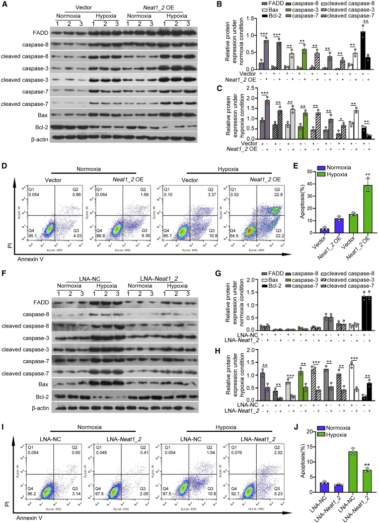
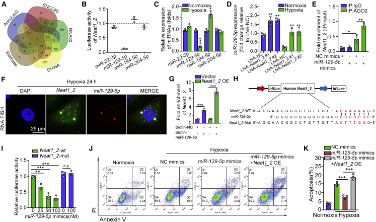
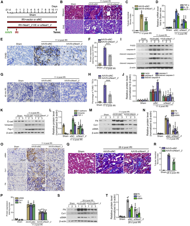
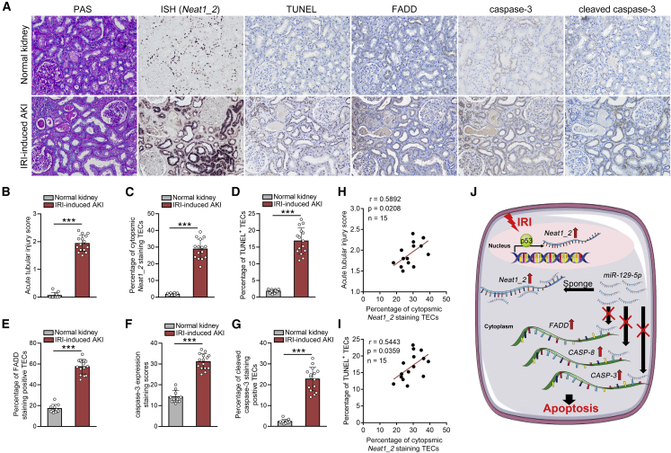
Acute kidney injury (AKI) is increasingly identified as a crucial risk factor for progression to CKD. However, the factors governing AKI to CKD progression remain largely unknown. By high-throughput RNA sequencing, we found that Neat1_2, a transcript variant of Neat1, was upregulated in 40-min ischemia/reperfusion injury (IRI), which resulted in the development of renal fibrotic lesions. The upregulation of Neat1_2 in hypoxia-treated TECs was attributed to p53 transcriptional regulation. Gain- and loss-of-function studies, both in vitro and in vivo, demonstrated that Neat1_2 promoted apoptosis of injured TECs induced by IRI and caused tubulointerstitial inflammation and fibrosis. Mechanistically, Neat1_2 shares miRNA response elements with FADD, CASP-8, and CASP-3. Neat1_2 competitively binds to miR-129-5p and prevents miR-129-5p from decreasing the levels of FADD, CASP-8, and CASP-3, and ultimately facilitates TEC apoptosis. Increased expression of Neat1_2 associated with kidney injury and TEC apoptosis was recapitulated in human AKI, highlighting its clinical relevance. These findings suggest that preventing TEC apoptosis by hindering Neat1_2 expression may be a potential therapeutic strategy for AKI to CKD progression.

Keywords: ESRD; acute kidney injury; apoptosis; chronic kidney disease; ischemic-reperfusion injury; long noncoding RNA; microRNA; renal fibrosis; tubular epithelial cell; tubulointerstitial fibrosis.


Conflict of interest statement

Declaration of interests The authors declare no competing interests.

Figures

None
Graphical abstract
Figure 1
Figure 1
LncRNA Neat1_2 is upregulated by p53 transcription in TECs from mice exposed to severe IRI (A) Clustered heatmap of the differentially expressed lncRNAs in isolated tubules from sham and IRI mice (n = 3). (B) Northern blots for the 2 transcript variants of Neat1 in isolated tubules from 20- or 40-min IRI mice (n = 3). (C) Expression of Neat1_2 in isolated tubules from sham and IRI mice, assayed by qPCR. (D and E) Expression of Neat1_2 in mouse tubular epithelial cells (mTECs) (D) and HK2 cells (E) treated with hypoxia (1% oxygen) for 24 h. (F) The subcellular location of Neat1_2 in normoxia or hypoxia-treated HK2 cells was analyzed by RNA-FISH. Scale bar, 25 μm. (G) Normoxia- or hypoxia-treated HK2 cells were analyzed for cytoplasmic and nuclear percentage of Neat1_2. (H and I) Representative images of in situ hybridization of Neat1_2 expression in kidneys from sham or IRI mice (H) and the percentage of cytoplasmic Neat1_2 staining TECs (I), Scale bars, 100 μm in the upper images and 30 μm in the lower images. (J) A Venn diagram showing potential transcription factors associated with Neat1_2. Transcription factors (p53 and Rela) were selected by using DNA pull down followed by liquid chromatography-tandem mass spectrometry as well as ChIPBase. (K) Knockdown of p53 decreased the Neat1_2 transcription activity in luciferase reporter assays. (L) ChIP assays showing p53 occupied at the Neat1_2 promoter in HK2 cells. (M) Luciferase reporter assays for HK2 cells that were transfected with reporter plasmids containing truncated Neat1_2 promoters and then treated with hypoxia for 24 h. (N) Mutation of p53 binding site rescued Neat1_2 transcription in luciferase reporter assays. The data are presented as means ± SDs. A Student’s t test was used for the comparison of 2 groups. ANOVA was used for comparison among multiple groups. ∗∗p < 0.01, ∗∗∗p < 0.001.
Figure 2
Figure 2
Upregulation of Neat1_2 activates TEC apoptosis (A–C) Representative western blot analyses and quantification data showed cellular expression of pro-apoptotic factors in Neat1_2-overexpressed HK2 cells in the presence or absence of hypoxia treatment for 24 h. (D and E) The effect of Neat1_2 overexpression on apoptosis and the quantification data. Cell apoptosis was assayed by co-staining of annexin V and propidium iodide followed by flow cytometric analysis. (F–H) Representative western blot analyses and quantification data showed cellular expression of pro-apoptotic factors in Neat1_2-knocked down HK2 cells in the presence or absence of hypoxia treatment for 24 h. (I and J) The effect of Neat1_2 knockdown on apoptosis and the quantification data. The data are presented as means ± SDs. A Student’s t test was used for the comparison of 2 groups. ANOVA was used for comparison among multiple groups. ∗p < 0.05, ∗∗p < 0.01, ∗∗∗p < 0.001.
Figure 3
Figure 3
Neat1_2 activates the apoptotic cascade via inhibition of miR-129-5p (A) Venn diagram of predicted miRNAs interacted with Neat1_2 in 5 databases. Four miRNAs were the intersection of these databases. (B) Luciferase activity of Neat1_2 in HEK-293T cells transfected with mimics of miRNAs that putatively bind to the Neat1_2 sequence. (C) Expression levels of the 4 putative Neat1_2-interacting miRNAs in HK2 cells treated with hypoxia for 24 h, assayed by qPCR. (D) Expression of the 4 putative Neat1_2-interacting miRNAs in HK2 cells transfected with locked nucleic acid (LNA) targeting Neat1_2 in the presence or absence of hypoxia treatment, assayed by qPCR. (E) RIP assay were performed using AGO2 antibody in HK2 cells transfected with miR-129-5p mimics or mimics negative control (mimic NC), then the enrichment of Neat1_2 was detected. (F) FISH was performed to observe the cellular location of Neat1_2 (green) and miR-129-5p (red) in HK2 cells treated with hypoxia for 24 h. Scale bar, 25 μm. (G) Neat1_2 was pulled down and enriched with 3′ end biotinylated miR-129-5p in the lysates of HK2 cells transfected with Neat1_2 overexpression plasmids or empty vector. (H) Schematic illustration of Neat1_2-WT and Neat1_2-Mut luciferase reporter vectors. (I) Luciferase assays in HEK-293T cells co-transfected with Neat1_2-WT or Neat1_2-Mut luciferase reporter vectors together with different doses of miR-129-5p mimics. (J and K) The effect of miR-129-5p mimics on apoptosis in HK2 cells treated with hypoxia for 24 h, in the presence or absence of Neat1_2 overexpression, and the quantification data. The data are presented as means ± SDs. A Student’s t test was used for the comparison of 2 groups. ANOVA was used for comparison among multiple groups. ∗p < 0.05, ∗∗p < 0.01, ∗∗∗p < 0.001.
Figure 4
Figure 4
miR-129-5p inhibits Neat1_2-mediated TEC apoptosis in vitro through targeting the 3′ UTR of FADD, CASP-3, and CASP-8 (A) Schematic illustration of FADD-WT, CASP-8-WT, CASP-3-WT, FADD-Mut, CASP-8-Mut, and CASP-3-Mut luciferase reporter vectors. (B-D) Luciferase assays in HEK-293T cells co-transfected with WT or mutated luciferase reporter vectors of FADD (B), CASP-8 (C), and CASP-3 (D), together with different doses of miR-129-5p mimics. (E) qPCR showing the expression of pro-apoptotic factors in HK2 cells transfected with mimic NC or miR-129-5p under hypoxia treatment for 24 h. (F and G) Western blot showing the expression of pro-apoptotic factors in HK2 cells transfected with mimic NC or miR-129-5p under hypoxia treatment for 24 h. (H) qPCR showing the expression of pro-apoptotic factors in Neat1_2-overexpressed HK2 cells transfected with mimic NC or miR-129-5p. (I and J) Western blot showing the expression of pro-apoptotic factors in Neat1_2-overexpressed HK2 cells transfected with mimic NC or miR-129-5p. (K) Luciferase assays in HK2 cells co-transfected with WT or mutated luciferase reporter vectors of FADD, CASP-8, and CASP-3, together with Neat1_2 depletion or overexpression. (L and M) The effects of miR-129-5p mimics on apoptosis in HK2 cells induced by Neat1_2 overexpression, and the quantification data. (N and O) Luciferase assays in HK2 cells co-transfected with luciferase reporter vectors of Bax and CASP-7 together with Neat1_2 overexpression. (P and Q) Western blot showing the expression of FADD, Bax, and caspase-7 in miR-129-5p inhibitor-transfected HK2 cells, together with FADD or scramble siRNAs transfection. The data are presented as means ± SDs. A Student’s t test was used for the comparison of 2 groups. ANOVA was used for comparison among multiple groups. ∗p < 0.05, ∗∗p < 0.01, ∗∗∗p < 0.001.
Figure 5
Figure 5
Knockdown of endogenous Neat1_2 protects against AKI after IRI by inhibiting TECs apoptosis (A) Mice were treated by renal vein injection of either AAV9-siNeat1_2 or AAV9-siNC 21 days before establishing IRI. (B and C). Representative images of ISH of Neat1_2 expression in kidneys from IRI mice injected with AAV9-siNeat1_2 (B) and the percentage of cytoplasmic Neat1_2 positive TECs (C). (D and E) qPCR showing the expression of Neat1_2 (D) and miR-129-5p (E) in renal cortex from IRI mice treated with AAV9-siNeat1_2. (F and G) Representative micrographs showing kidney injury in different groups 1 day after IRI injury (F), and the quantification data of acute tubular injury score (G). (H) Serum creatinine level in IRI mice injected with either AAV9-siNeat1_2 or AAV9-siNC 1 day post IRI. (I and J) Representative images of TUNEL staining showing a reduced percentage of TECs apoptosis in the kidneys from IRI mice injected with AAV9-siNeat1_2 (I), and the quantification data (J). (K) qPCR showed that depletion of Neat1_2 decreased the mRNA expression of pro-apoptotic factors in renal cortex homogenates from mice subjected to IRI. (L and M) Representative immunohistochemistry images showing the expression levels of pro-apoptotic factors in IRI mice injected with either AAV9-siNeat1_2 or AAV9-siNC 1 day post IRI (L), and the quantification data (M). (N and O) Western blot showed that depletion of Neat1_2 decreased the protein expression of pro-apoptotic factors in renal cortex homogenates from mice subjected to IRI. Scale bars in (B), (F), (I), (L) are 200 μm. The data are presented as means ± SDs. A Student’s t test was used for the comparison of 2 groups. ANOVA was used for comparison among multiple groups (n = 6 for each group). ∗∗∗p < 0.001.
Figure 6
Figure 6
Expression of exogenous Neat1_2 aggravates AKI after IRI (A) Mice were treated by renal vein injection of either AAV9-Vector or AAV9-Neat1_2 21 days before performing IRI. (B and C) Representative images of ISH of Neat1_2 expression in kidneys from IRI mice injected with AAV9-Neat1_2 (B) and the percentage of cytoplasmic Neat1_2 positive TECs (C). (D and E) qPCR showing the expression of Neat1_2 (D) and miR-129-5p (E) in renal cortex from IRI mice treated with AAV9-Neat1_2. (F) Serum creatinine level in IRI mice injected with either AAV9-Vector or AAV9-Neat1_2 1 day post IRI. (G and H) Representative images showing kidney injury in different groups 1 day after IRI injury (G), and the quantification data of acute kidney injury score (H). (I and J) Representative images of TUNEL staining showing an increased TECs apoptosis in the kidneys from IRI mice injected with AAV9-Neat1_2 (I), and the quantification data (J). (K) qPCR showed that ectopic expression of Neat1_2 further increased the mRNA expression of pro-apoptotic factors in kidney homogenates from mice subjected to IRI. (L and M) Western blot showed that overexpression of Neat1_2 in IRI mice further promoted the expression of pro-apoptotic factors in kidney homogenates from mice subjected to IRI. (N and O) Representative images of immunohistochemistry showed that overexpression of Neat1_2 in IRI mice further promoted the expression of pro-apoptotic factors. Scale bars in (B), (G), (I), (N) are 200 μm. The data are presented as means ± SDs. A Student’s t test was used for the comparison of 2 groups. ANOVA was used for comparison among multiple groups (n = 6 for each group). ∗∗∗p < 0.001.
Figure 7
Figure 7
Inhibition of Neat1_2 preserves tubular integrity and improves tubulointerstitial fibrosis (A) Mice were treated by renal vein injection of AAV9 to manipulate the expression of Neat1_2 in mouse kidney 21 days before performing IRI. Mice were sacrificed 11 days post-injury. (B and C) Representative images of Masson’s trichrome staining in mice treated with either AAV9-siNeat1_2 or AAV9-siNC (B), and the quantification data (C). (D) qPCR showed that depletion of Neat1_2 decreased the mRNA expression of pro-inflammatory factors in kidney homogenates from mice subjected to IRI. (E and F) Representative images of immunohistochemical staining of F4/80 in kidney tissue from mice treated with either AAV9-siNeat1_2 or AAV9-siNC 11 days post-injury. (G and H) Representative images and quantification data of TUNEL staining showed that depletion of Neat1_2 inhibited TEC apoptosis. (I and J) Western blot showed that depletion of Neat1_2 in IRI mice alleviated the expression of pro-apoptotic factors in kidney homogenates from mice subjected to IRI 11 days post-injury. (K and L) Western blot showed that depletion of Neat1_2 in IRI mice preserved tubular integrity, as reflected by the increase in E-cadherin and decrease in vimentin and FSP-1. (M and N) Western blot showed that inhibition of Neat1_2 expression decreased the expression of profibrotic factors 11 days post-IRI. (O and P) Immunohistochemical staining of profibrotic factors in kidney tissue from mice treated with either AAV9-siNeat1_2 or AAV9-siNC 11 days post-injury. (Q and R) Representative images and quantification data of Masson’s trichrome staining in IRI mice injected with either AAV9-siNC or AAV9-siNeat1_2 28 days post-IRI treatment. (S and T) Western blot showed that inhibition of Neat1_2 expression decreased the expression of profibrotic factors 28 days post-IRI. Scale bars in (B), (E), (G), (Q) are 200 μm. Scale bar in (O) is 100 μm. The data are presented as means ± SDs. A Student’s t test was used for the comparison of 2 groups. ANOVA was used for comparison among multiple groups (n = 6 for each group). ∗∗∗p < 0.001.
Figure 8
Figure 8
Neat1_2 upregulation correlates with epithelial cell apoptosis in human AKI (A–G) Representative images of ISH for Neat1_2 and TUNEL staining and immunohistochemistry for pro-apoptotic factors in human kidney sections from patients with AKI (A) and quantification data (n = 10 for control, n = 15 for patients with AKI) (B–G). Scale bar in (A) is 200 μm. (H and I) Correlation between Neat1_2 expression and acute tubular injury score (H), or percentage of TUNEL staining positive TECs (I). (J) Graphical abstract of this study. The data are presented as means ± SDs. In (A)–(G), p values were calculated by Student’s t test. In (H) and (I), p values were calculated by Pearson correlation analysis.

Similar articles

Cited by

References

    1. Kalantar-Zadeh K., Jafar T.H., Nitsch D., Neuen B.L., Perkovic V. Chronic kidney disease. Lancet. 2021;398:786–802. doi: 10.1016/s0140-6736(21)00519-5. - DOI - PubMed
    1. Liu Y. Renal fibrosis: new insights into the pathogenesis and therapeutics. Kidney Int. 2006;69:213–217. doi: 10.1038/sj.ki.5000054. - DOI - PubMed
    1. Chawla L.S., Eggers P.W., Star R.A., Kimmel P.L. Acute kidney injury and chronic kidney disease as interconnected syndromes. N. Engl. J. Med. 2014;371:58–66. doi: 10.1056/NEJMra1214243. - DOI - PMC - PubMed
    1. Ishani A., Xue J.L., Himmelfarb J., Eggers P.W., Kimmel P.L., Molitoris B.A., Collins A.J. Acute kidney injury increases risk of ESRD among elderly. J. Am. Soc. Nephrol. 2009;20:223–228. doi: 10.1681/asn.2007080837. - DOI - PMC - PubMed
    1. Wald R., Quinn R.R., Luo J., Li P., Scales D.C., Mamdani M.M., Ray J.G. Chronic dialysis and death among survivors of acute kidney injury requiring dialysis. Jama. 2009;302:1179. doi: 10.1001/jama.2009.1322. - DOI - PubMed

Publication types

MeSH terms