. 2022 May 10:13:815041.
doi: 10.3389/fimmu.2022.815041. eCollection 2022.

Skewed Cellular Distribution and Low Activation of Functional T-Cell Responses in SARS-CoV-2 Non-Seroconvertors

Affiliations

Skewed Cellular Distribution and Low Activation of Functional T-Cell Responses in SARS-CoV-2 Non-Seroconvertors

Athina Kilpeläinen et al. Front Immunol. .

Abstract

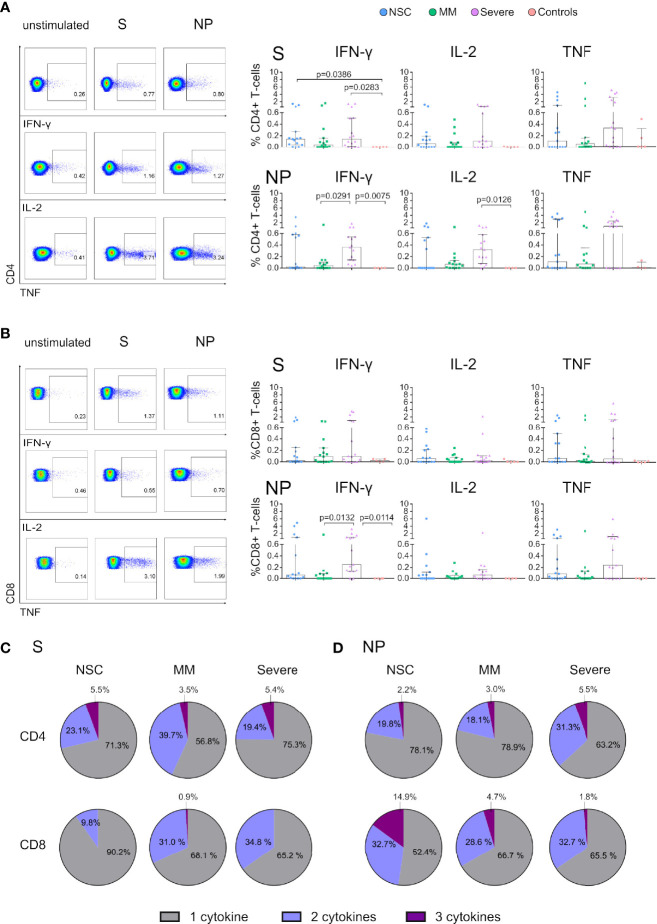
The role of T cells in the control of SARS-CoV-2 infection has been underestimated in favor of neutralizing antibodies. However, cellular immunity is essential for long-term viral control and protection from disease severity. To understand T-cell immunity in the absence of antibody generation we focused on a group of SARS-CoV-2 Non-Seroconvertors (NSC) recovered from infection. We performed an immune comparative analysis of SARS-CoV-2 infected individuals stratified by the absence or presence of seroconversion and disease severity. We report high levels of total naïve and low effector CD8+ T cells in NSC. Moreover, reduced levels of T-cell activation monitored by PD-1 and activation-induced markers were observed in the context of functional SARS-CoV-2 T-cell responses. Longitudinal data indicate the stability of the NSC phenotype over three months of follow-up after infection. Together, these data characterized distinctive immunological traits in NSC including skewed cellular distribution, low activation and functional SARS-CoV-2 T-cell responses. This data highlights the value of T-cell immune monitoring in populations with low seroconversion rates in response to SARS-CoV-2 infection and vaccination.

Keywords: SARS-CoV-2; T cell subsets; cellular immunity; function; immune activation; non-seroconvertor.


Conflict of interest statement

The authors declare that the research was conducted in the absence of any commercial or financial relationships that could be construed as a potential conflict of interest.

Figures

Figure 1
Figure 1
CD4+ and CD8+ T-cell subset distribution in Non-Seroconvertors. (A) Flow chart of the study groups, including Non-Seroconvertors (NSC), Mild-moderate (MM), Severe (B, C) Cryopreserved PBMCs were cultured and surface stained with antibodies targeting CD4 and CD8 as well as T-cell lineage markers CD45RA, CCR7, CD27 to distinguish T-central memory cells (TCM), T effector memory cells (TEM), T transitional memory cells (TTM), T Naïve cells (TN) and T effector cells (TEF). Cells were pre-gated for lymphocytes and live cells. Whisker plots show the frequencies of CD4+ T-cell subsets and pie charts represent the frequency distribution of CD4+ and CD8+ T-cell subsets in each group (Panel B, C, respectively). Statistical analyses were performed using a multivariable regression analysis. Only significant values are indicated in the Figure. Linear regression was performed plotting relative frequencies of CD8+ TN (D) and CD8+ TEF (E) against the age of the study participants, as well as relative frequencies of CD8+ TN (F) and CD8+ TEF (G) against the Days from Symptom Onset (DfSO) to sample. Correlation analyses were performed using Spearman’s rank correlation test; r- and p-values are reported for each group.
Figure 2
Figure 2
Expression of activation markers is lower in SARS-CoV-2 Non-Seroconvertors compared to seropositive individuals. PBMCs from Non-Seroconvertors and comparative study groups were stimulated for 17 hours with S and NP recombinant proteins and stained using antibodies directed against activation-induced markers (AIM; CD25, OX40, CD137) and PD-1, and analysed by flow cytometric analysis. Cells were pre-gated for lymphocytes and live cells. (A) Representative dot-plots of AIM expression in CD4+ and CD8+ T cells. (B) Bar graphs showing the expression of activation induced markers in CD4+ (CD25+/OX40+) and CD8+ T cells in response to S and NP (CD137+). Values from unstimulated cells were subtracted for each individual. (C) Representative dot-plots of PD-1 expression on CD4+ and CD8+ T cells. (D) Bar graphs of the expression of PD-1 on CD4+ and CD8+ T cells in unstimulated cells as well as following S and NP stimulation. Error bars represent the interquartile range and median values per group are shown. Statistical analysis was performed using Kruskal-Wallis non-parametric ANOVA adjusted for multiple comparisons, only significant differences are highlighted in the Figure.
Figure 3
Figure 3
Functional T cells of SARS-CoV-2 Non-Seroconvertors are present in NSC in response to Spike and Nucleocapsid. Cryopreserved PBMCs from SARS-CoV-2 Non-Seroconvertors (NSC) and comparative study groups, Mild-moderate (MM), Severe, and controls samples were stimulated for 17 hours with Spike (S) and Nucleocapsid proteins (NP). An unstimulated condition was used as a control and values were subtracted. Cells were pre-gated for lymphocytes and live cells. Production of IFN-γ, IL-2, and TNF was monitored by Flow cytometric analysis following intracellular cytokine staining. (A, B) Left panels depict the gating strategy for analysis of cytokine expression and right panels represent the frequencies of IFN-γ, IL-2, and TNF in CD4+ (A) and CD8 (B) T cells in the study groups in response to S and NP. Pie charts demonstrate the relative proportions of T cells producing one, two or three cytokines in response to S in CD4+ T cells and CD8+ T cells (C). Relative proportions of T cells producing one, two or three cytokines in response to NP in CD4+ T cells and CD8+ T cells (D). Statistical analysis was performed using non-parametric ANOVA (Kruskal Wallis) adjusted for multiple comparisons. Error bars represent the interquartile range and median values per group are shown. Only significant differences are indicated in the Figure.

References

    1. Chang AY, Cullen MR, Harrington RA, Barry M. The Impact of Novel Coronavirus COVID-19 on Noncommunicable Disease Patients and Health Systems: A Review. J Intern Med (2021) 289(4):450–62. doi: 10.1111/joim.13184 - DOI - PMC - PubMed
    1. Blanco-Melo D, Nilsson-Payant BE, Liu WC, Uhl S, Hoagland D, Møller R, et al. . Imbalanced Host Response to SARS-CoV-2 Drives Development of COVID-19. Cell (2020) 181:1036–45.e9. doi: 10.1016/j.cell.2020.04.026 - DOI - PMC - PubMed
    1. Marshall M. The Lasting Misery of Coronavirus Long-Haulers. Nature (2020) 585:339–41. doi: 10.1038/d41586-020-02598-6 - DOI - PubMed
    1. Li T, Xie J, He Y, Fan H, Baril L, Qiu Z, et al. . Long-Term Persistence of Robust Antibody and Cytotoxic T Cell Responses in Recovered Patients Infected With SARS Coronavirus. PloS One (2006) 1(1):e24.doi: 10.1371/journal.pone.0000024 - DOI - PMC - PubMed
    1. Dan JM, Mateus J, Kato Y, Hastie KM, Yu ED, Faliti CE, et al. . Immunological Memory to SARS-CoV-2 Assessed for Up to 8 Months After Infection. Science (80- ) (2021) 371(6529):eabf4063. doi: 10.1126/science.abf4063 - DOI - PMC - PubMed

Publication types