. 2022 May 10:13:829406.
doi: 10.3389/fimmu.2022.829406. eCollection 2022.

Tolerance of a Vascularized Composite Allograft Achieved in MHC Class-I-mismatch Swine via Mixed Chimerism

Affiliations

Tolerance of a Vascularized Composite Allograft Achieved in MHC Class-I-mismatch Swine via Mixed Chimerism

Alexandre G Lellouch et al. Front Immunol. .

Abstract

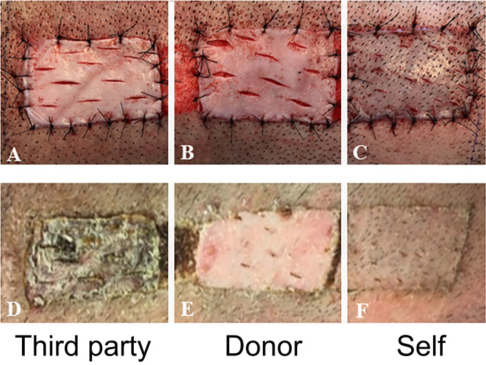
Background: Vascularized composite allografts (VCAs) allow reconstruction of devastating injuries and amputations, yet require lifelong immunosuppression that is associated with significant morbidity. Induction of immune tolerance of VCAs would permit widespread use of these procedures. VCAs are acquired from deceased donors most likely to be fully-MHC-mismatched (in contrast to living-related renal transplant donor-recipient pairs matched at one MHC haplotype). After achieving VCA tolerance in a swine model equivalent to clinical living-related renal transplants (single-haplotype MHC mismatches: e.g., "mother-daughter"/haploidentical), we tested our protocol in MHC class I, class II, and fully-MHC-mismatched pairs. Although class II mismatched swine demonstrated similar results as the haploidentical scenario (stable mixed chimerism and tolerance), our protocol failed to prevent rejection of class I and full mismatch VCAs. Here, we describe a new adapted conditioning protocol that successfully achieved tolerance across MHC class-I-mismatch barriers in swine.

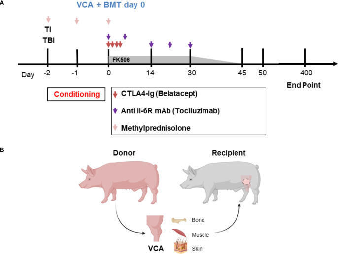
Methods: Swine were treated with non-myeloablative total body and thymic irradiation two days prior to infusion of bone marrow cells from an MHC class I-mismatched donor. They also received a short-term treatment with CTLA4-Ig (Belatacept®) and anti-IL6R mAb (Tociluzimab®) and were transplanted with an osteomyocutaneous VCA from the same donor.

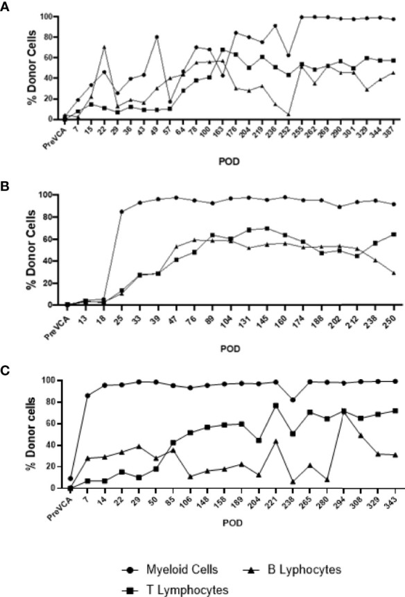
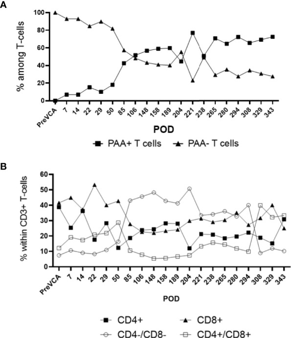
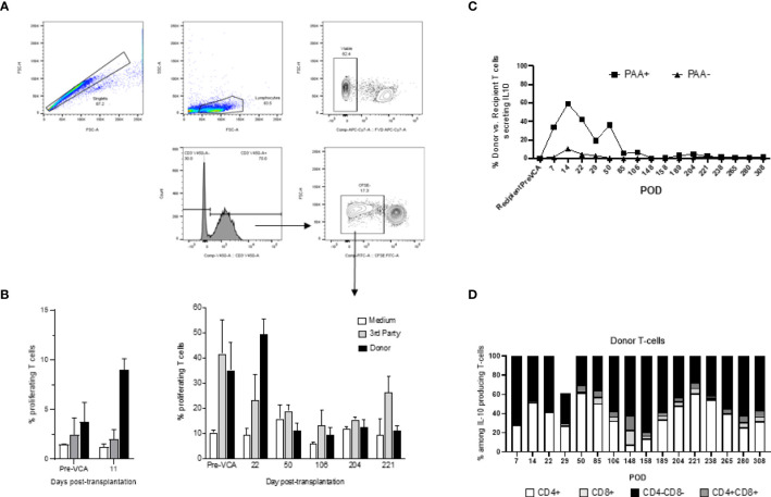
Results: Stable mixed chimerism and tolerance of MHC class-I-mismatched VCAs was achieved in 3 recipients. Allograft tolerance was associated with a sustained lack of anti-donor T cell response and a concomitant expansion of double negative CD4-CD8- T cells producing IL-10.

Conclusions: This study demonstrates the first successful mixed chimerism-induced VCA tolerance in a large animal model across a MHC class-I-mismatch. Future studies aimed at fully-mismatched donor-recipient pairs are under investigation with this protocol.

Keywords: MHC class I; Vascularized composite allotransplantation (VCA); bone marrow transplantation; co-stimulatory blockade; mixed chimerism; skin tolerance.


Conflict of interest statement

The authors declare that the research was conducted in the absence of any commercial or financial relationships that could be construed as a potential conflict of interest.

Figures

Figure 1
Figure 1
(A) Mixed chimerism protocol for immune tolerance. Recipient conditioning begins prior to transplantation with total body irradiation (TBI, day -2) and thymic irradiation (TI, day -1). Induction is performed with methylprednisolone (days -2, -1, 0) and FK506. Following vascularized composite allotransplantation (VCA), the donor is exsanguinated, and bone marrow is harvested for transplantation (BMT) under the cover of CTLA4-Ig (Belatacept®) and anti-IL6R mAb (Tociluzimab®). FK506 is then continued for 30 days (target trough levels: 10-15 ng/mL) before gradual tapering to discontinuation on day 45; further CTLA4-Ig (Belatacept®) and anti-IL6R mAb (Tociluzimab®) is given I.V. 20 mg/kg (on POD 2, 4, 6) and I.V. 10 mg/kg (on POD 7, 14, 21, 28) respectively. (B) Schematic of donor hindlimb VCA tissue components and placement in the recipient. Image created with BioRender.
Figure 2
Figure 2
Clinical assessment of successful VCA tolerance. Images of tolerant VCAs from R3 (A), R4 (C), and R5 (E) taken on POD 251, 387, and 250, respectively, compared to a representative rejected VCA from untreated control R6 on POD 11 (G). H&E staining of VCA biopsies from R3 (B), R4 (D), and R5 (F), taken on POD 151, 387, and 250 respectively, demonstrating absence of rejection (Banff Grade 0). Whereas H&E staining of VCA skin from class I mismatch untreated control R6 shows focal epidermal necrosis and capillary thrombosis (Banff Grade IV) on POD 11 (H). (POD, post-operative day; VCA, vascularized composite allograft).
Figure 3
Figure 3
Long-term multi-lineage mixed chimerism in recipient animals R4 (A), R3 (B) and R5 (C). Levels of donor chimerism in myeloid cells were >80%, whereas B and T cell populations ranged between 30 to 60%.
Figure 4
Figure 4
Sub-analysis of T cell subsets in multi-lineage mixed chimerism. Post-transplantation, as bone marrow reconstitution progresses, the percentage of donor (PAA+) and recipient (PAA-) T cells approaches equilibrium at around POD100, remained stable and varied between 50 to 70% of donor T cells to experimental endpoint (A). The population of double negative (CD4-/CD8-) T cells increase from around POD 50 onwards (following cessation of FK506) and remained at about 20 to 40% of overall CD3+ T cell to experimental endpoint (B). The results shown were obtained in one tolerant swine and are representative of all tolerant swine tested individually.
Figure 5
Figure 5
In vivo test of immune competence by placement of split-thickness skin grafts from a third-party animal (A), original donor (B), and self (C) at POD 150 onto recipient swine tolerant of VCA (representative). By POD 14, there was complete rejection of the skin graft from the third-party animal (D) whereas that from the donor (E) and self (F) had taken.
Figure 6
Figure 6
(A) Gating strategy for mixed lymphocyte reaction (MLR) assay. Cells analyzed in this assay were first gated as singlet events, followed by gating on the major lymphocyte population. Fixable viability dye negative events (viable) were analyzed for CD3+ expression. This population was characterized based on CFSE fluorescence, where proliferating T-cells were denoted as CFSE dim and plotted as a proportion of total CD3+ events. (B) Mixed lymphocyte reaction assay testing proliferation of T cells in vitro (left panel untreated controls, right panel tolerant chimeric recipients). Untreated control animals demonstrate increased T-cell proliferation against donor targets at time of rejection (left panel), whereas tolerant animals have decreased T-cell responsiveness towards the donor overtime (right panel). From POD 50 onwards, which corresponds more or less to the withdrawal of all immunosuppression and establishment of multi-lineage mixed chimerism, cellular proliferation against donor and self-antigens were comparable to medium, suggesting a state of immune unresponsiveness in peripheral blood (right panel). (C) Similar to Figure 3 which showed equilibration of donor (PAA+) and recipient (PAA-) contribution to overall T cell populations, the contribution of IL-10 secreting T cells followed a similar pattern, approaching equilibrium at POD 85. Further analysis revealed that the majority of such IL-10 producing T cells are CD4-CD8- in nature (D). The results shown in panel (C, D) were obtained in one tolerant swine and are representative of all tolerant swine tested individually.

References

    1. Dubernard JM, Owen E, Herzberg G, Lanzetta M, Martin X, Kapila H, et al. Human Hand Allograft: Report on First 6 Months. Lancet (1999) 353(9161):1315–20. doi: 10.1016/s0140-6736(99)02062-0 - DOI - PubMed
    1. Rifkin WJ, Manjunath AK, Kantar RS, Jacoby A, Kimberly LL, Gelb BE, et al. A Comparison of Immunosuppression Regimens in Hand, Face, and Kidney Transplantation. J Surg Res (2021) 258:17–22. doi: 10.1016/j.jss.2020.08.006 - DOI - PubMed
    1. Brannstrom M, Diaz-Garcia C, Johannesson L, Dahm-Kahler P, Bokstrom H. Livebirth After Uterus Transplantation - Authors' Reply. Lancet (2015) 385(9985):2352–3. doi: 10.1016/S0140-6736(15)61098-4 - DOI - PubMed
    1. Petruzzo P, Sardu C, Lanzetta M, Dubernard JM. Report, (2017) of the International Registry on Hand and Composite Tissue Allotransplantation (IRHCTT). Curr Transplant Rep (2017) 4:294–303. doi: 10.1007/s40472-017-0168-3 - DOI
    1. Conrad A, Petruzzo P, Kanitakis J, Gazarian A, Badet L, Thaunat O, et al. Infections After Upper Extremity Allotransplantation: A Worldwide Population Cohort Study 1998-2017. Transpl Int (2019) 32(7):693–701. doi: 10.1111/tri.13399 - DOI - PubMed

Publication types