AQP5 Is a Novel Prognostic Biomarker in Pancreatic Adenocarcinoma
- PMID: 35619903
- PMCID: PMC9128544
- DOI: 10.3389/fonc.2022.890193
AQP5 Is a Novel Prognostic Biomarker in Pancreatic Adenocarcinoma
Abstract
Background: Pancreatic adenocarcinoma (PAAD) is a highly malignant tumor with a poor prognosis. The identification of effective molecular markers is of great significance for diagnosis and treatment. Aquaporins (AQPs) are a family of water channel proteins that exhibit several properties and play regulatory roles in human carcinogenesis. However, the association between Aquaporin-5 (AQP5) expression and prognosis and tumor-infiltrating lymphocytes in PAAD has not been reported.
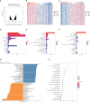
Methods: AQP5 mRNA expression, methylation, and protein expression data in PAAD were analyzed using GEPIA, UALCAN, HAP, METHSURV, and UCSC databases. AQP5 expression in PAAD patients and cell lines from our cohort was examined using immunohistochemistry and Western blotting. The LinkedOmics database was used to study signaling pathways related to AQP5 expression. TIMER and TISIDB were used to analyze correlations among AQP5, tumor-infiltrating immune cells, and immunomodulators. Survival was analyzed using TCGA and Kaplan-Meier Plotter databases.
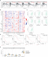
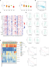
Results: In this study, we investigated AQP5 expression in PAAD and determined whether the expression of AQP5 is a strong prognostic biomarker for PAAD. We searched and analyzed public cancer databases (GEO, TCGA, HAP, UALCAN, GEPIA, etc.) to conclude that AQP5 expression levels were upregulated in PAAD. Kaplan-Meier curve analysis showed that high AQP5 expression positively correlated with poor prognosis. Using TIMER and TISIDB, we found that the expression of AQP5 was associated with different tumor-infiltrating immune cells, especially macrophages. We found that hypomethylation of the AQP5 promoter region was responsible for its high expression in PAAD.
Conclusions: AQP5 can serve as a novel biomarker to predict prognosis and immune infiltration in PAAD.
Keywords: AQP5; methylation; pancreatic adenocarcinoma; prognosis; tumor microenvironment.
Copyright © 2022 Chen, Song, Yang, Du, Zheng, Lu, Zhang and Wei.
Conflict of interest statement
The authors declare that the research was conducted in the absence of any commercial or financial relationships that could be construed as a potential conflict of interest.
Figures


Similar articles
-
High expression of RNF169 is associated with poor prognosis in pancreatic adenocarcinoma by regulating tumour immune infiltration.Front Genet. 2023 Jan 5;13:1022626. doi: 10.3389/fgene.2022.1022626. eCollection 2022. Front Genet. 2023. PMID: 36685833 Free PMC article.
-
A Comprehensive Pan-Cancer Analysis Reveals GRB7 as a Potential Diagnostic and Prognostic Biomarker.Cureus. 2024 Dec 1;16(12):e74907. doi: 10.7759/cureus.74907. eCollection 2024 Dec. Cureus. 2024. PMID: 39742197 Free PMC article.
-
Unraveling the role of Major Vault Protein as a novel immune-related biomarker that promotes the proliferation and migration in pancreatic adenocarcinoma.Front Immunol. 2024 Jul 4;15:1399222. doi: 10.3389/fimmu.2024.1399222. eCollection 2024. Front Immunol. 2024. PMID: 39026679 Free PMC article.
-
Comprehensive analysis of the expression and prognostic value of ARMCs in pancreatic adenocarcinoma.BMC Cancer. 2025 Jan 7;25(1):28. doi: 10.1186/s12885-024-13365-5. BMC Cancer. 2025. PMID: 39773340 Free PMC article.
-
Identify potential prognostic indicators and tumor-infiltrating immune cells in pancreatic adenocarcinoma.Biosci Rep. 2022 Feb 25;42(2):BSR20212523. doi: 10.1042/BSR20212523. Biosci Rep. 2022. PMID: 35083488 Free PMC article. Review.
Cited by
-
Evaluation of tumor targets selected from public genomic databases for imaging of pancreatic ductal adenocarcinoma.Sci Rep. 2025 May 16;15(1):17102. doi: 10.1038/s41598-025-00517-1. Sci Rep. 2025. PMID: 40379788 Free PMC article.
-
Aquaporin-5 Dynamic Regulation.Int J Mol Sci. 2023 Jan 18;24(3):1889. doi: 10.3390/ijms24031889. Int J Mol Sci. 2023. PMID: 36768212 Free PMC article. Review.
-
Overexpression of OAS1 Is Correlated With Poor Prognosis in Pancreatic Cancer.Front Oncol. 2022 Jul 11;12:944194. doi: 10.3389/fonc.2022.944194. eCollection 2022. Front Oncol. 2022. PMID: 35898870 Free PMC article.
-
Clinical diagnosis and management of pancreatic cancer: Markers, molecular mechanisms, and treatment options.World J Gastroenterol. 2022 Dec 28;28(48):6827-6845. doi: 10.3748/wjg.v28.i48.6827. World J Gastroenterol. 2022. PMID: 36632312 Free PMC article. Review.
-
Expression of aquaporin 1, 3 and 5 in colorectal carcinoma: correlation with clinicopathological characteristics and prognosis.Pathol Oncol Res. 2023 Jun 2;29:1611179. doi: 10.3389/pore.2023.1611179. eCollection 2023. Pathol Oncol Res. 2023. PMID: 37334171 Free PMC article.
References
LinkOut - more resources
Full Text Sources

