Review
. 2022 May 10:16:871223.
doi: 10.3389/fnint.2022.871223. eCollection 2022.

Unexpected Consequences of Noise-Induced Hearing Loss: Impaired Hippocampal Neurogenesis, Memory, and Stress

Affiliations
Review

Unexpected Consequences of Noise-Induced Hearing Loss: Impaired Hippocampal Neurogenesis, Memory, and Stress

Senthilvelan Manohar et al. Front Integr Neurosci. .

Abstract

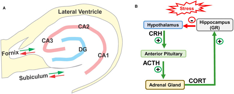
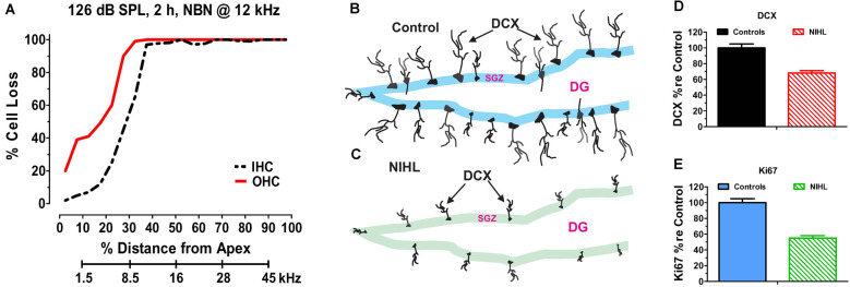
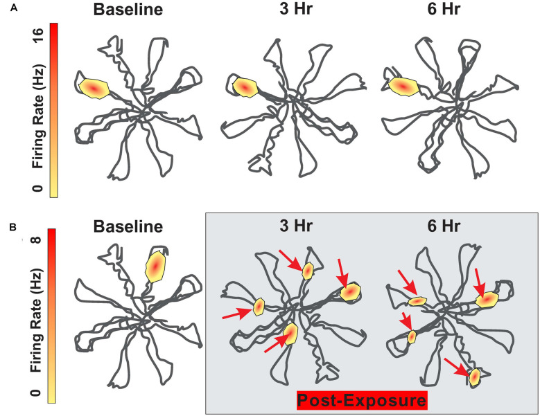
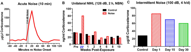
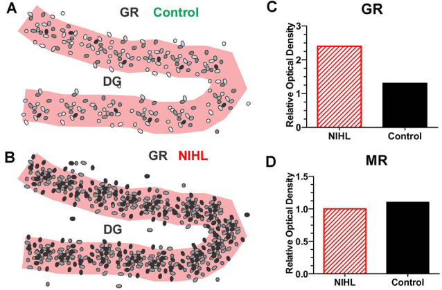
Noise-induced hearing loss (NIHL), caused by direct damage to the cochlea, reduces the flow of auditory information to the central nervous system, depriving higher order structures, such as the hippocampus with vital sensory information needed to carry out complex, higher order functions. Although the hippocampus lies outside the classical auditory pathway, it nevertheless receives acoustic information that influence its activity. Here we review recent results that illustrate how NIHL and other types of cochlear hearing loss disrupt hippocampal function. The hippocampus, which continues to generate new neurons (neurogenesis) in adulthood, plays an important role in spatial navigation, memory, and emotion. The hippocampus, which contains place cells that respond when a subject enters a specific location in the environment, integrates information from multiple sensory systems, including the auditory system, to develop cognitive spatial maps to aid in navigation. Acute exposure to intense noise disrupts the place-specific firing patterns of hippocampal neurons, "spatially disorienting" the cells for days. More traumatic sound exposures that result in permanent NIHL chronically suppresses cell proliferation and neurogenesis in the hippocampus; these structural changes are associated with long-term spatial memory deficits. Hippocampal neurons, which contain numerous glucocorticoid hormone receptors, are part of a complex feedback network connected to the hypothalamic-pituitary (HPA) axis. Chronic exposure to intense intermittent noise results in prolonged stress which can cause a persistent increase in corticosterone, a rodent stress hormone known to suppress neurogenesis. In contrast, a single intense noise exposure sufficient to cause permanent hearing loss produces only a transient increase in corticosterone hormone. Although basal corticosterone levels return to normal after the noise exposure, glucocorticoid receptors (GRs) in the hippocampus remain chronically elevated. Thus, NIHL disrupts negative feedback from the hippocampus to the HPA axis which regulates the release of corticosterone. Preclinical studies suggest that the noise-induced changes in hippocampal place cells, neurogenesis, spatial memory, and glucocorticoid receptors may be ameliorated by therapeutic interventions that reduce oxidative stress and inflammation. These experimental results may provide new insights on why hearing loss is a risk factor for cognitive decline and suggest methods for preventing this decline.

Keywords: glucocorticoid receptor (GCR); hippocampus; memory; neurogenesis; noise-induced hearing loss; spatial navigation; stress.


Conflict of interest statement

The authors declare that the research was conducted in the absence of any commercial or financial relationships that could be construed as a potential conflict of interest.

Figures

Figure 1
Figure 1
(A) Schematic of hippocampus and surrounding lateral ventricle. Three subdivisions of the rodent Cornu Ammonis (CA1, CA2, and CA3) surrounding the dentate gyrus (DG). Only the major afferent and efferent pathways (red/green arrows) through the subiculum and fornix are shown. (B) Schematic of hypothalamic-pituitary adrenal (HPA) axis with hippocampus. Stress stimulates the release (+) of corticotropic releasing hormone (CRH) from the hypothalamus, which binds to receptors in anterior pituitary causing the release (+) of adrenocorticotropic hormone (ACTH) which stimulates the release of corticosterone (CORT). CORT binds to glucocorticoid receptors (GRs) in the hippocampus, which provides negative feedback (−) to the hypothalamus suppressing the release of CRH, ACTH, and CORT.
Figure 2
Figure 2
Noise-inducedhearing loss suppresses hippocampal cell proliferation andneurogenesis. (A) Cochleogram showing massive loss of outerhair cells (OHC) and inner hair cells (IHC) in the noise-exposed cochlear several months after a 2-h unilateral exposure to narrowband noise (NBN) centered at 12 kHz and presented at 126 dB SPL. Percent cell loss plotted as function of percent distance from the apex of the cochlear. Cochlear place related to frequency using rat tonotopic map on lower abscissa. (B) Schematic of dentate gyrus (DG) of hippocampus from normal control showing immunolabeled doublecortin (DCX) soma in the subgranular zone (SGZ); note extensive immunolabeled processes emanating from soma. (C) Schematic of DG of hippocampus several months after a noised induced hearing loss (NIHL) showing immunolabeled DCX) soma in the subgranular zone (SGZ). Note reduced number of DCX soma and paucity of labeled processes in the NIHL hippocampus compared to normal control (panel B). (D) Schematic showing relative number (% re Control: percentage relative to control) of DCX labeled neurons in hippocampus of normal control rats (100%) and rats with noise-induced hearing loss (NIHL). (E) Schematic showing relative number (% re Control: percentage relative to control) of Ki67 labeled neurons in hippocampus of normal control rats (100%) and rats with noise-induced hearing loss (NIHL).
Figure 3
Figure 3
Schematic showing firing pattern of place cells in the hippocampus as rat navigates through an eight-arm radial maze. (A) Schematic showing place cell firing pattern within the radial maze; place at which the cell fires remain relatively stable between baseline and 3 h and 6 h later. (B) Schematic showing place cell firing pattern at baseline and then 3 h and 6 h following 30 min exposure to 104 dB SPL 4 kHz tone. Location of place cell firing locations drastically altered after the noise exposure. Maximum firing rate on upper and lower heat maps is 16 Hz and 8 Hz respectively.
Figure 4
Figure 4
Noise-induced changes in corticosterone. (A) Schematic showing rapid rise and fall of serum corticosterone following a 10-min exposure to white noise presented at 114 dB SPL. (B) Schematic illustrating corticosterone levels measured over 12 weeks in sham control rats and rats exposed unilaterally for 2-h to 126 dB SPL narrowband noise (NBN) centered at 12 kHz. Corticosterone greatly elevated in noise group 20’ post-exposure, but levels decline to normal 1-week post-exposure. No significant difference in long-term basal corticosterone levels between control and noise-exposed group. (C) Schematic illustrating the rise in serum corticosterone following chronic intermittent noise presented at 100 dB SPL for 4-h/day over a period of 30 days. On day 1, corticosterone measured 30-min post-exposure while on day 15 and day 30, corticosterone was measured 24-h after the exposure.
Figure 5
Figure 5
Severe NIHL alters glucocorticoid receptor (GR) expression in hippocampus. (A) Schematic of dentate gyrus (DG) in hippocampus showing immunolabeling of GR receptors (black, gray round, oval symbols schematically illustrate the relative intensity of immunolabeling.) in normal control. (B) Schematic of DG in hippocampus showing GR immunolabeling several months after induction of severe unilateral noise-induced hearing loss (NIHL; 126 dB SPL, 2 h, NBN centered at 12 kHz). (C) Schematic of relative optical density of GR immunolabeling in DG in rats with severe chronic NIHL compared to controls. (D) Schematic of relative optical density of mineralocorticoid receptor (MR) immunolabeling of rats with chronic NIHL compared to controls.

References

    1. Alzheimer’s Association Report. (2020). 2020 Alzheimer’s disease facts and figures. Alzheimers Dement. 10.1002/alz.12068 - DOI - PubMed
    1. Adlard P. A., Perreau V. M., Engesser-Cesar C., Cotman C. W. (2004). The timecourse of induction of brain-derived neurotrophic factor mRNA and protein in the rat hippocampus following voluntary exercise. Neurosci. Lett. 363, 43–48. 10.1016/j.neulet.2004.03.058 - DOI - PubMed
    1. Adlard P. A., Perreau V. M., Pop V., Cotman C. W. (2005). Voluntary exercise decreases amyloid load in a transgenic model of Alzheimer’s disease. J. Neurosci. 25, 4217–4221. 10.1523/JNEUROSCI.0496-05.2005 - DOI - PMC - PubMed
    1. Agasse F., Mendez-David I., Christaller W., Carpentier R., Braz B. Y., David D. J., et al. . (2020). Chronic corticosterone elevation suppresses adult hippocampal neurogenesis by hyperphosphorylating huntingtin. Cell Rep. 32:107865. 10.1016/j.celrep.2020.107865 - DOI - PubMed
    1. Agnihotri N. T., Hawkins R. D., Kandel E. R., Kentros C. (2004). The long-term stability of new hippocampal place fields requires new protein synthesis. Proc. Natl. Acad. Sci. U S A 101, 3656–3661. 10.1073/pnas.0400385101 - DOI - PMC - PubMed

LinkOut - more resources