Review
. 2022 Mar 22;8(1):42-53.
doi: 10.1002/wjo2.12. eCollection 2022 Mar.

Three-hundred and sixty degrees of surgical approaches to the maxillary sinus

Affiliations
Review

Three-hundred and sixty degrees of surgical approaches to the maxillary sinus

Natália C Rezende et al. World J Otorhinolaryngol Head Neck Surg. .

Abstract

Objectives: To demonstrate three-hundred and sixty degrees of maxillary sinus (MS) surgical approaches using cadaveric dissections, highlighting the step-by-step anatomy of each procedure.

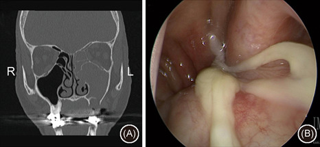
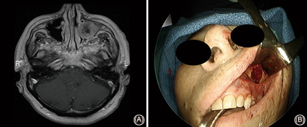
Methods: Two latex-injected cadaveric specimens were utilized to perform surgical dissections to demonstrate different approaches to the MS. The procedures were documented with macroscopic images and endoscopic pictures.

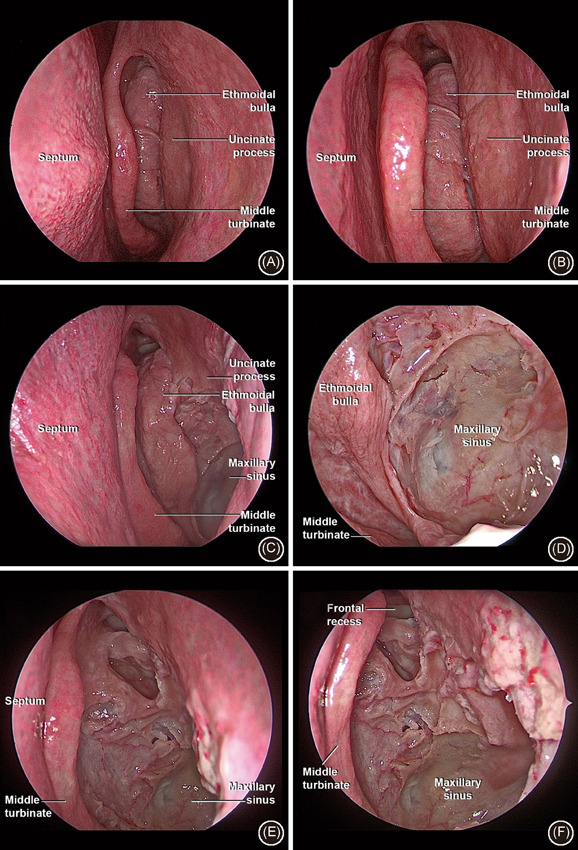
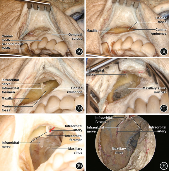
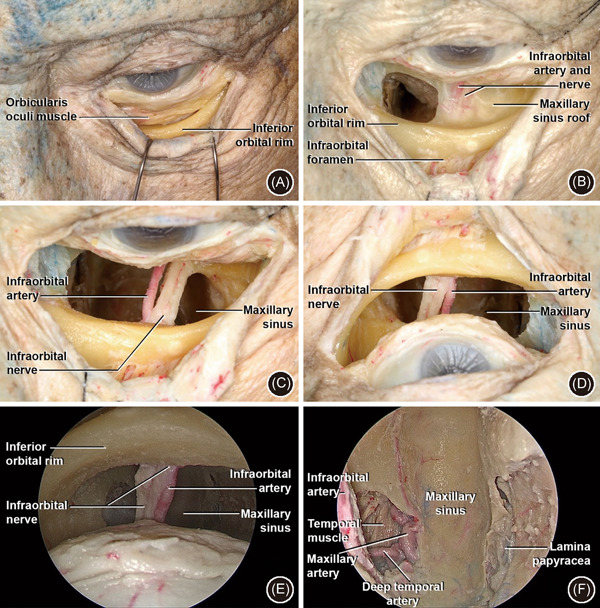
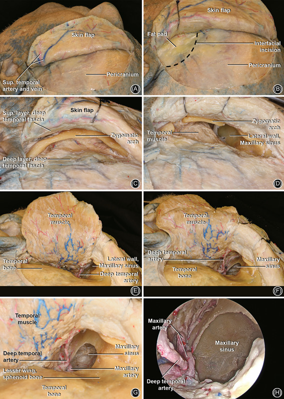
Results: Dissections were performed to approach the MS medially (endoscopic maxillary antrostomy and ethmoidectomy), anteriorly (Caldwell-Luc), superiorly (transconjunctival/transorbital approach), inferiorly (transpalatal approach), and posterolaterally (preauricular hemicoronal approach).

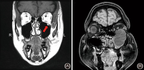
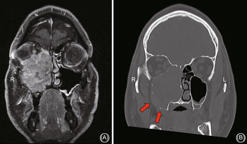
Conclusion: A number of approaches have been described to address pathology in the MS. Surgeons should be familiar with indications, limitations, and surgical anatomy from different perspectives to approach the MS. This paper illustrates anatomic approaches to the MS with detailed step-by-step cadaveric dissections and case examples.

Keywords: Caldwell–Luc; anatomy; dissection; endoscopic sinus surgery; infratemporal fossa; maxillary sinus; pterygopalatine fossa.


Conflict of interest statement

The authors declare that there are no conflict of interests.

Figures

Figure 1
Figure 1
Endoscopic anterior ethmoidectomy (antrostomy) approach. (A) Endoscopic overview of the left nasal cavity and middle meatus; (B) medialization of middle turbinate with better visualization of ethmoid bulla; (C) immediately after completion of uncinectomy and initial expansion of the antrostomy; (D) further enlargement of the antrostomy posteriorly to connect the natural and any accessory ostium of MS; (E) complete removal of bulla; (F) overview of final aspect of maxillary sinus anterior ethmoidectomy
Figure 2
Figure 2
Case example of utilization of maxillary antrostomy and anterior ethmoidectomy for a left‐sided odontogenic sinusitis; (A) coronal noncontrast CT scan demonstrating left‐sided maxillary and anterior ethmoid sinus opacification adjacent to a bony defect on the sinus floor with periapiacal abscess and (b) endoscopic exam demonstrating purulence emanating from the middle meatus and draining into the nasopharynx. CT, computed tomography
Figure 3
Figure 3
Caldwell–Luc (anterior transmaxillary) approach. (A) Right side: incision between the canine tooth and second molar, about 5 mm above gingival sulcus; (B) detachment of periosteum of maxilla and identification of canine fossa and eminence inferior and medially; (C) identification of IOF and ION in the central‐superior area; (D) window in anterior wall (3 cm diameter) with visualization of maxillary sinus mucosa. ION in central‐superior area; (E) removal of sinus mucosa and opening into the MS cavity. Visualization of ION and IOA; (F) 4 mm 0‐degree endoscope MS anterior view with ION and IOA in the posterior wall. IOA, infraorbital artery; IOF, infraorbital foramen; ION, infraorbital nerve; MS, maxillary sinus
Figure 4
Figure 4
Case example of utilization of a Caldwell–Luc approach; left‐sided maxillary sinus squamous cell carcinoma requiring a combined endoscopic medial maxillectomy and Caldwell–Luc approach; (A) axial T1 postgadolinium MRI scan showing left‐sided maxillary sinus tumor and (B) intraoperative photo demonstrating Caldwell–Luc approach through the anterior wall of the maxillary sinus. MRI, magnetic resonance imaging
Figure 5
Figure 5
Transconjunctival approach. (A) Left side: conjunctival incision just below to lower border of the tarsus with detachment between septum and orbicularis oculi muscle in the orbital rim; (B) subperiosteal detachment of orbital floor and identification of ION and IOA. The IOF with ION and IOA are identified in the anterior surface of the inferior orbital rim. A window has been opened in the orbit floor medial to the ION; (C) the bone has been drilled medial and lateral to the ION and IOA—anatomic view; (D) surgical view; of C (E): 0‐degree endoscopic superior view of MS, with ION and IOA—surgical view; (F): 0 degree endoscopic superior view of MS, with IMAX and DTA laterally (on the left) and papyracea lamina medially (on the right)—surgical view. DTA, deep temporal artery; IMAX, internal maxillary artery; IOA, infraorbital artery; IOF, infraorbital foramen; ION, infraorbital nerve; MS, maxillary sinus
Figure 6
Figure 6
Case examples of pathology requiring a transconjunctival approach; (A) coronal T1 postgadolinium MRI scan demonstrating left infraorbital nerve enlargement and hyperintensity from perineural invasion from cutaneous malignancy (red arrow) and (B) coronal T1 postgadolinium MRI scan demonstrating a large left‐sided V2 schwannoma requiring a combined endoscopic and Caldwell–Luc approach for resection and adjuvant transconjunctival approach for orbital floor reconstruction. MRI, magnetic resonance imaging
Figure 7
Figure 7
Transpalatal approach. (A) Overview of the oral cavity with hard palate visualization and inverted “U” incision in left hemipalatal region. (B) Mucoperiosteal flap with hard palate bone exposure. Greater palatine artery in inferior/lateral portion of hard palate; (C) a burr hole has been performed in the hard palate with an inferior view of the MS, mucosal flap, and GPA; (D) 4 mm 0‐degree endoscopic inferior overview of MS with ION in the posterior wall. GPA, great palatine artery; ION, infraorbital nerve; MS, maxillary sinus
Figure 8
Figure 8
Case example of a right‐sided maxillary sinus mucosal melanoma requiring a combined transpalatal and transfacial approach with orbital exenteration; (A) coronal T1 postgadolinium MRI scan showing intraorbital extension and (B) coronal noncontrast CT scan demonstrating bony erosion and tumor involvement along the floor of sinus necessitating transpalatal approach. CT, computed tomography; MRI, magnetic resonance imaging
Figure 9
Figure 9
Preauricular hemicoronal approach. (A) Left side: incision 1–2 cm posterior to the hairline extending from the preauricular area towards the forehead midline. (B) Raise the skin flap above the periosteal layer. An incision is performed at the level of the fat pad (black dashed line) 3–4 cm above supraorbital ridge, and dissection is carried out anteriorly in an interfascial plane to expose the zygoma. The superficial layer of the deep temporal fascia is kept with the skin flap to protect the frontal branches of the facial nerve that runs superficial to the superficial layer of the deep temporal fascia. (C) Overview of the zygoma and the superficial layer of the deep temporal fascia superiorly; (D) osteotomies in the lateral and medial portion of the zygomatic arch; (E) elevation of the temporal muscle flap and visualization of posterior/lateral wall of MS. Note the DTA; (F) open window in the lateral wall of MS. (G) Enlarged view of (F), note the DTA and IMAX; (H) 0 degree endoscopic lateral view of MS. DTA, deep temporal artery; IMAX, internal maxillary artery; MS, maxillary sinus
Figure 10
Figure 10
Case examples of pathology necessitating a preauricular hemicoronal approach; (A) noncontrast coronal CT scan of extensive maxillofacial trauma including zygomatic complex fractures and (B) axial T1 postgadolinium MRI scan of sinonasal squamous cell carcinoma requiring a combined craniotomy and hemicoronal approach. CT, computed tomography; MRI, magnetic resonance imaging

References

    1. Whyte A, Boeddinghaus R. The maxillary sinus: physiology, development and imaging anatomy. Dentomaxillofac Radiol. 2019;48:20190205. - PMC - PubMed
    1. Cho DY, Hwang PH. Results of endoscopic maxillary mega‐antrostomy in recalcitrant maxillary sinusitis. Am J Rhinol. 2008;22:658‐662. - PubMed
    1. Woo HJ, Hwang PH, Kaplan MJ, Choby G. Clinical characteristics and prognostic factors of malignant tumors involving pterygopalatine fossa. Head Neck. 2020;42:281‐288. - PubMed
    1. Wang EW, Gullung JL, Schlosser RJ. Modified endoscopic medial maxillectomy for recalcitrant chronic maxillary sinusitis. Int Forum Allergy Rhinol. 2011;1:493‐497. - PubMed
    1. Peng R, Thamboo A, Choby G, Ma Y, Zhou B, Hwang PH. Outcomes of sinonasal inverted papilloma resection by surgical approach: an updated systematic review and meta‐analysis. Int Forum Allergy Rhinol. 2019;9:573‐581. - PubMed