

Cutaneous fistula on the cheek associated with oculo-auriculo-vertebral spectrum
- PMID: 35620227
- PMCID: PMC9129258
- DOI: 10.1093/jscr/rjac187
Cutaneous fistula on the cheek associated with oculo-auriculo-vertebral spectrum
Abstract
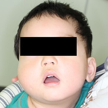
A Japanese boy, presented with epibulbar dermoid and ipsilateral preauricular appendages, had a pit on his cheek of the same side. An atrial septal defect and vertebral fusions were also identified. He was diagnosed with a mild type of oculo-auriculo-vertebral spectrum (OAVS). At the age of 18 months, his cheek was swollen with a slight fever. An infected cyst and cutaneous fistula enveloped by the risorius muscle were extracted. It was assumed to be a remnant of the fissure between the maxillary and mandibular prominences. This was the first case of cutaneous fistula confirmed histologically with OAVS, although there seem to be more cases. The possibility of the mechanism of smiling cheek dimple is also discussed.
Published by Oxford University Press and JSCR Publishing Ltd. © The Author(s) 2022.
Figures


References
-
- Rollnick BR, Kaye CI, Nagatoshi K, Hauck W, Martin AO. Oculoauriculovertebral dysplasia and variants: phenotypic characteristics of 294 patients. Am J Med Genet 1987;26:361–75. - PubMed
-
- Allen T. A description of congenital malformation of the auricle and external meatus of both sides in three persons, with experiments on the state of hearing in them, and remarks on the mode of hearing by conduction through the hard parts of the head in general. N Proc R Soc Edinburgh 1845;1:443–6.
-
- Grabb WC. The first and second branchial arch syndrome. Plast Reconstr Surg 1965;36:485–508. - PubMed
-
- Finn DG, Buchalter IH, Sarti E, Romo T. First branchial cleft cysts: clinical update. Laryngoscope 1987;97:136–40. - PubMed
-
- Alasti F, Van Camp G. Genetics of microtia and associated syndromes. J Med Genet 2009;46:361–9. - PubMed
Publication types
LinkOut - more resources
Full Text Sources
