. 2022 May 6;25(6):104364.
doi: 10.1016/j.isci.2022.104364. eCollection 2022 Jun 17.

Altered synaptic plasticity of the longitudinal dentate gyrus network in noise-induced anxiety

Affiliations

Altered synaptic plasticity of the longitudinal dentate gyrus network in noise-induced anxiety

Sojeong Pak et al. iScience. .

Abstract

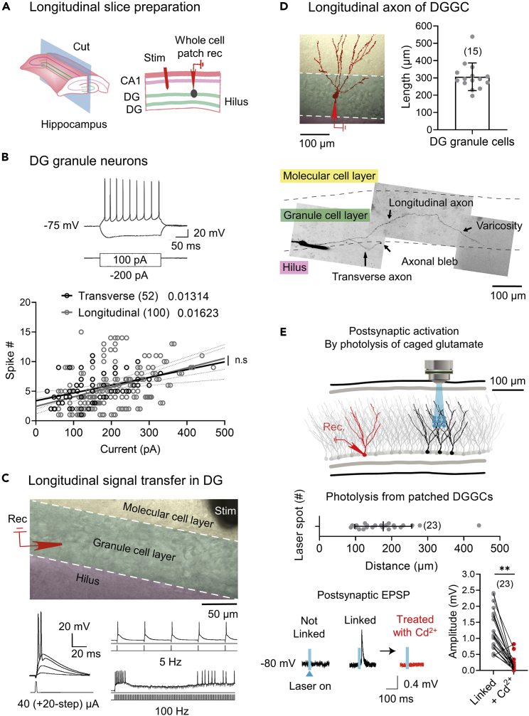
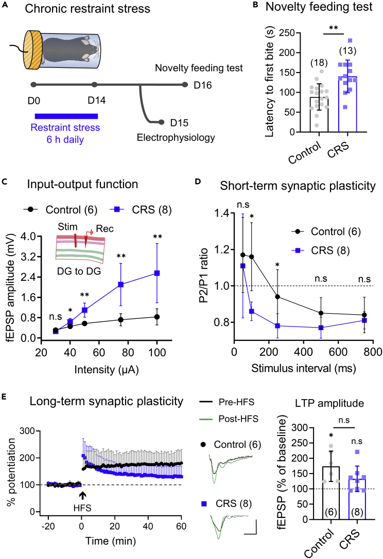
Anxiety is characteristic comorbidity of noise-induced hearing loss (NIHL), which causes physiological changes within the dentate gyrus (DG), a subfield of the hippocampus that modulates anxiety. However, which DG circuit underlies hearing loss-induced anxiety remains unknown. We utilize an NIHL mouse model to investigate short- and long-term synaptic plasticity in DG networks. The recently discovered longitudinal DG-DG network is a collateral of DG neurons synaptically connected with neighboring DG neurons and displays robust synaptic efficacy and plasticity. Furthermore, animals with NIHL demonstrate increased anxiety-like behaviors similar to a response to chronic restraint stress. These behaviors are concurrent with enhanced synaptic responsiveness and suppressed short- and long-term synaptic plasticity in the longitudinal DG-DG network but not in the transverse DG-CA3 connection. These findings suggest that DG-related anxiety is typified by synaptic alteration in the longitudinal DG-DG network.

Keywords: Behavioral neuroscience; Biological sciences; Neuroscience.


Conflict of interest statement

The authors declare no competing interests.

Figures

None
Graphical abstract
Figure 1
Figure 1
NIHL enhances anxious behaviors (A) Timeline of the experimental procedure. (B) Hearing threshold (dB sound pressure level [SPL]) at different frequency tones, as measured by the auditory brainstem response (ABR) recorded during pre- and post-noise (D7) exposure. Noise exposure elevates the hearing threshold. (C) The total number of entries and the latency to the first entry into each arm in the Y-maze forced alteration task. NIHL significantly increases the latency to the first entry to the novel arm. (D) The total distance traveled in an open field arena and the exploration time with the novel or familiar object during object recognition. Both the control and NIHL have novel object preference over familiar one. (E) The total area moved and the time (s) ratio of the center zone to the outer zone in the open field test. The ratio is reduced in NIHL. Individual data points have been eliminated for clearer visibility. (F) The latency to the first food bite in the novelty-suppressed feeding test. In NIHL, the latency to food biting is increased. The numbers in parentheses indicate the number of animals tested. Data are presented as means ± SD. NIHL: noise-induced hearing loss; ∗p < 0.05; ∗∗p < 0.01; n.s.: not significant. Also, see Table S1.
Figure 2
Figure 2
NIHL minimally alters the DG-CA3 network (A) Hippocampal transverse slice preparation and configurations of the recording (Rec) and stimulating (Stim) electrodes in the slice used to examine the synaptic properties of the DG-CA3 network. (B) The input-output function of CA3, represented as the fEPSP amplitude in response to increasing electrical stimulation of the DG granule cell layer, showed no significant differences between the control and NIHL groups. (C) Short-term synaptic plasticity in response to a paired-pulse stimulation at various inter-pulse intervals. The P2/P1 (pulse2/pulse1) ratio is not significantly different between the control and NIHL groups. (D) Long-term synaptic potentiation (LTP) at the DG-CA3 synapses, is induced by theta-burst stimulation (TBS). The insets display sample fEPSP traces of pre (black)- and post (green)-TBS induction (the scale bar denotes 5 ms × 0.2 mV). DCG-IV (mGluR2/3 agonist) is treated at the end of LTP experiment. The LTP amplitude is significantly increased compared to the normalized baseline (100%) in both groups and is not significantly different between the control and NIHL groups. The numbers in parentheses indicate the number of brain slices tested. Data are presented as means ± SD. NIHL, noise-induced hearing loss; DG, dentate gyrus; fEPSP, field excitatory postsynaptic potential; ∗p < 0.05; ∗∗p < 0.01; n.s, not significant. Also, see Tables S2 and S3.
Figure 3
Figure 3
Longitudinal slicing angle enables signals to propagate longitudinally in the DG network (A) Hippocampal longitudinal slice preparation and configuration of the stimulating (Stim) and recording (Rec) electrode and for whole-cell patch recording in the slice. (B) DGGCs exhibit regular firing patterns in response to depolarizing current injection. The relationship between the number of spikes and the depolarizing current injection is similarly linear in both transverse and longitudinal slices. The floating numbers are positive progression slopes. (C) The configurations of the recording and stimulating electrodes in hippocampal longitudinal slices when examining signal flow from the DG-DG network in the presence of picrotoxin (50 μM). The whole cell-patched DGGC generates sub-threshold responses and action potentials in response to increasing intensities of electrical stimulation in the molecular cell layer. The 5-Hz stimulation successfully induces spikes, and the 100-Hz stimulation induces refractory periods in the middle of the spike trains. (D) The somatic and dendritic morphology of a DGGC, and a reconstructed image of a recorded DGGC filled with Alexa Fluor 594 showing axonal projections to the granule cell layer (a longitudinal axon) and to the hilus (a transverse axon). (E) Glutamate uncaging is facilitated by local dot-photostimulation (cyan circle) of DGGCs that are hundreds of microns away from a patched cell (red) within the DG layer. Depolarization is induced in a postsynaptic patched neuron when it is linked to a presynaptic photo-activated neuron. Glutamate-induced EPSPs are far reduced in the presence of Cd2+, which blocks synaptic transmission. The numbers in parentheses indicate the number of cells tested. Data are presented as means ± SD. DG, dentate gyrus; DGGC, dentate gyrus granule cell; EPSP, excitatory postsynaptic potential; ∗∗ p < 0.01.
Figure 4
Figure 4
Comparison of the synaptic properties of the DG-DG networks in transverse and longitudinal slices of the hippocampus (A) A schematic of the transverse and longitudinal slices of the hippocampus. Both longitudinal and transverse slices are prepared with stimulating (Stim) and recording (Rec) electrodes positioned near the borders of the molecular and granule cell layers. The input-output function of DG, represented as the fEPSP amplitude in response to increasing electrical stimulation of the DG granule cell layer, showed higher synaptic responsiveness in longitudinal slices than in transverse slices. (B) Short-term synaptic plasticity in response to a paired-pulse stimulation at various inter-pulse intervals. The P2/P1 (pulse2/pulse1) ratio is not significantly different between the transverse and longitudinal slices. (C) Long-term synaptic potentiation (LTP) in both preparations, is tested by high-frequency stimulation (HFS). The insets display sample fEPSP traces of pre (black)- and post (red)-HFS induction. The LTP amplitude in the longitudinal slices is significantly increased compared to the normalized baseline (100%) and is significantly different between the transverse and longitudinal slices. The numbers in parentheses indicate the number of brain slices tested. Data are presented as means ± SD. DG, dentate gyrus; fEPSP, field excitatory postsynaptic potential; ∗∗p < 0.01. Also, see Tables S2 and S3.
Figure 5
Figure 5
NIHL suppresses synaptic plasticity in the longitudinal DG-DG network (A) Hippocampal longitudinal slice preparation and configurations of the stimulating (Stim) and recording (Rec) electrodes in the slice used to examine the synaptic properties of the DG-DG network. (B) The input-output function of DG, represented as the fEPSP amplitude in response to increasing electrical stimulation of the inner molecular layer of the DG, shows hyperexcitable responses in the NIHL group. (C) Long-term synaptic potentiation (LTP) at the DG-DG synapses, is induced by high-frequency stimulation (HFS). The insets display sample fEPSP traces of pre (black)- and post (green)-HFS induction (The scale bar denotes 5 ms × 0.2 mV). The LTP amplitude in the control group is significantly increased compared to the normalized baseline (100%) but not in the NIHL group and is significantly different between the control and NIHL groups. NIHL eliminates LTPs in the longitudinal DG network. (D) Short-term synaptic plasticity in response to a paired-pulse stimulation at various inter-pulse intervals. The NIHL condition shows paired-pulse depression and is significantly more depressed than the control group in the P2/P1 (pulse2/pulse1) ratio. (E) Immunohistochemistry to detect neuronal viability in the hippocampal DG, stained with an antibody against NeuN (green) and a stain of nuclear DNA (DAPI, blue). The relative NeuN integrated density (NeuN intensity normalized to the DAPI intensity) indicates no significant neuronal cell loss in the DG after NIHL. The numbers in parentheses indicate the number of brain slices tested. Data are presented as means ± SD. NIHL: noise-induced hearing loss; DG, dentate gyrus; fEPSP, field excitatory postsynaptic potential; ∗p < 0.05; ∗∗p < 0.01; n.s, not significant. Also, see Tables S2 and S3.
Figure 6
Figure 6
Stress-induced anxiety alters synaptic plasticity in the longitudinal DG network (A) Schematic of a chronic stress-induced anxiety model and timeline of the experimental procedure. (B) The latency to the first food bite in the novelty-suppressed feeding test. Chronic restraint stress (CRS) mice show increased latency to the first food bite and display anxiety-like behaviors. The numbers in parentheses indicate the number of animals tested. (C) The input-output function of DG, represented as the fEPSP amplitude in response to increasing electrical stimulation of the inner molecular layer of the DG, shows hyperexcitable responses in the CRS group. The insert shows a hippocampal longitudinal slice and the configurations of the stimulating (Stim) and recording (Rec) electrodes when examining the synaptic properties of the DG-DG network. (D) Short-term synaptic plasticity in response to a paired-pulse stimulation at various inter-pulse intervals. The CRS condition shows paired-pulse depression and is significantly more depressed than the control group in the P2/P1 (pulse2/pulse1) ratio. (E) Long-term synaptic potentiation (LTP) at the DG-DG synapses, is induced by high-frequency stimulation (HFS). The insets display sample fEPSP traces of pre (black)- and post (green)-HFS induction (The scale bar denotes 5 ms × 0.2 mV). The LTP amplitude in the control group is significantly increased compared to the normalized baseline (100%) but not in the CRS group and is not significantly different between the control and CRS groups. CRS impairs LTPs in the longitudinal network. The numbers in parentheses indicate the number of brain slices tested. Data are presented as means ± SD. DG, dentate gyrus; fEPSP, field excitatory postsynaptic potential; ∗p < 0.05; ∗∗p < 0.01; n.s: not significant. Also, see Tables S2 and S3.

References

    1. Amaral D.G., Scharfman H.E., Lavenex P. The dentate gyrus: fundamental neuroanatomical organization (dentate gyrus for dummies) Prog. Brain Res. 2007;163:3–790. doi: 10.1016/s0079-6123(07)63001-5. - DOI - PMC - PubMed
    1. Anacker C., Luna V.M., Stevens G.S., Millette A., Shores R., Jimenez J.C., Chen B., Hen R. Hippocampal neurogenesis confers stress resilience by inhibiting the ventral dentate gyrus. Nature. 2018;559:98–102. doi: 10.1038/s41586-018-0262-4. - DOI - PMC - PubMed
    1. Bhatt J.M., Bhattacharyya N., Lin H.W. Relationships between tinnitus and the prevalence of anxiety and depression. Laryngoscope. 2017;127:466–469. doi: 10.1002/lary.26107. - DOI - PMC - PubMed
    1. Blaabjerg M., Zimmer J. The dentate mossy fibers: structural organization, development and plasticity. Prog. Brain Res. 2007;163:85–803. doi: 10.1016/s0079-6123(07)63005-2. - DOI - PubMed
    1. Chiba S., Numakawa T., Ninomiya M., Richards M.C., Wakabayashi C., Kunugi H. Chronic restraint stress causes anxiety-and depression-like behaviors, downregulates glucocorticoid receptor expression, and attenuates glutamate release induced by brain-derived neurotrophic factor in the prefrontal cortex. Prog. Neuropsychopharmacol. Biol. Psychiatry. 2012;39:112–119. doi: 10.1016/j.pnpbp.2012.05.018. - DOI - PubMed

LinkOut - more resources