Review
. 2022 Aug;41(6):1316-1322.
doi: 10.1002/nau.24963. Epub 2022 May 27.

Surgical anatomy of the vaginal vault

Affiliations
Review

Surgical anatomy of the vaginal vault

Bernard T Haylen et al. Neurourol Urodyn. 2022 Aug.

Abstract

Aim: Vaginal vault (VV) surgery should be a key part of surgery for a majority of pelvic organ prolapse (POP). The surgical anatomy of the VV, the upper most part of the vagina, has not been recently subject to a dedicated examination and description.

Methods: Cadaver studies were performed in (i) 10 unembalmed cadaveric pelves (observation); (ii) 2 unembalmed cadaveric pelves (dissection); (iii) 5 formalinized hemipelves (dissection). The structural outline and ligamentous supports of the VV were determined. Further confirmation of observations in post-hysterectomy patients were from a separate study on 300 consecutive POP repairs, 46% of whom had undergone prior hysterectomy.

Results: The VV is equivalent to the Level I section of the vagina, measured posteriorly from the top of the posterior vaginal wall (apex or highest part of the vagina) to 2.5 cm below this point. It comprises the anterior fornix (through which cervix protrudes or is removed at hysterectomy), posterior fornix and two lateral fornices. Before hysterectomy, the posterior aspects of the cervix and upper vagina are supported by the uterosacral (USL) and cardinal ligaments (CL), the distal segments of which fuse together to form a cardinal-uterosacral ligament complex (cardinal utero-sacral complex), around 2-3 cm long. Post---hysterectomy, there is some residual USL support to the anterior fornix but the posterior fornix has no ligamentous support and is thus more vulnerable to prolapse.

Conclusion: Effective management of VV prolapse will need to be part of most POP repairs. Enhanced understanding of the surgical anatomy of the vaginal vault allows more effective planning of those POP surgeries.

Keywords: Level I Vagina; cysto-enterocele; pelvic organ prolapse; recto-enterocele; surgical anatomy; vaginal vault.


Conflict of interest statement

The authors declare no conflicts of interest.

Figures

Figure 1
Figure 1
(A) Vaginal vault is the Level I section of the vagina (apex to 2.5 cm below this in posterior measurement), ; (B) axial view of VV before hysterectomy; (C) axial view of VV posthysterectomy. VV, vaginal vault
Figure 2
Figure 2
(A) (above—diagrammatic) (B) (below—cadaver): Cardinal ligament (CL) and Uterosacral (USL) have their distal segments fuse together to create a Cardinal utero‐sacral complex (CUSC). From the cervix VV, the CL and passes postero‐laterally whilst the USL passes supero‐posteriorly. CL, cardinal ligaments; VV, vaginal vault
Figure 3
Figure 3
The distal segments of the USL and CL are fused. They are cut with the first pedicle of a vaginal hysterectomy, with CL more laterally placed. CL, cardinal ligaments; USL, uterosacral
Figure 4
Figure 4
USL, CL and round ligaments displayed in the one axial cadaveric cut. (Visible Human Project, National Library of Medicine, NLM. USA—with permission). CL, cardinal ligaments; USL, uterosacral
Figure 5
Figure 5
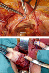
(A–C): (Short version). The intermediate section of the USL becomes visible, at the lower end of an anterior repair, just above the cuff scar line (A) when it is put under tension (B). Sutures can be used (C) to plicate the USL (Green—5): (Longer version) (A) A midline anterior vaginal wall incision with bladder mobilization and retraction. No USL is apparent as a wide shallow horizontal needle pass occurs through the dorsolateral aspect of the exposed vaginal vault superior to the cuff. (B) The intermediate section of the USL is readily seen when placed under tension. (C) Small inset shows the midline anterior vaginal wall incision (Inc) over the bladder (Bl) with the clamp (Cl) positioned to invert the vaginal vault to the vaginal introitus. The main part of (C) is a diagrammatic representation of (B) showing, 1, right‐angled retractor; 2, retracted bladder; 3, retrovesical space; 4, inverted vaginal vault; and 5, USL placed under tension at the beginning of plication. CL, cardinal ligaments; USL, uterosacral
Figure 6
Figure 6
(A, left): Total posterior vaginal length (TPVL). (B, right): Distance from vaginal vault (on traction) to anterior perineum. Posterior vaginal vault descent (PVVD) is the subtraction of the latter measurement from the TPVL
Figure 7
Figure 7
(A) Anterior‐oblique view of posthysterectomy posterior vagina (anterior to the rectum) with cuff scar (marked) anterior to apex. (B) Apparent “rectocele” though with cuff scar at top of “bulge”, this makes it a recto‐enterocele. (C) By pushing back postero‐superiorly with forceps, the combined prolapse (vault and posterior) disappears, replicating a VV repair. Thus, the majority of the posterior prolapse is VV descent. VV, vaginal vault
Figure 8
Figure 8
(A) Anterior‐oblique view of posthysterectomy: bladder, vagina, rectum; (B) Large cysto‐enterocele—bladder behind anterior vaginal wall (marked) and cuff scar (marked). The cysto‐enterocele with components of cystocoele and VV descent. The cuff scar is seen towards the bottom of the cystocele. VV, vaginal vault

References

    1. Summers A, Winkel LA, Hussain HK, DeLancey JOL. The relation between anterior and apical compartment support. Am J Obstet Gynecol. 2006;194:1438‐1443. - PMC - PubMed
    1. Haylen BT, Naidoo S, Kerr SJ, Chiu HJ, Birrell W. Posterior vaginal compartment repairs: where are the main anatomical defects? Int Urogynecol J. 2016;27:741‐745. - PubMed
    1. Haylen BT, Maher CF, Barber MD, et al. An International Urogynecological Association (IUGA)/International Continence Society (ICS) Joint report on the Terminology for Pelvic Organ Prolapse. Dual publication. Int Urogynecol J. 2016;27(2):165‐194. - PubMed
    1. DeLancey JOL. Anatomical aspects of vaginal eversion after hysterectomy. Am J Obst Gynecol. 1992;166:117‐124. - PubMed
    1. Zemlyn S. The length of the uterine cervix and its significance. J Clin Ultrasound. 1981;9(6):267‐269. - PubMed