. 2022 May 19;9(5):219.
doi: 10.3390/bioengineering9050219.

Human-Origin iPSC-Based Recellularization of Decellularized Whole Rat Livers

Affiliations

Human-Origin iPSC-Based Recellularization of Decellularized Whole Rat Livers

Aylin Acun et al. Bioengineering (Basel). .

Abstract

End-stage liver diseases lead to mortality of millions of patients, as the only treatment available is liver transplantation and donor scarcity means that patients have to wait long periods before receiving a new liver. In order to minimize donor organ scarcity, a promising bioengineering approach is to decellularize livers that do not qualify for transplantation. Through decellularization, these organs can be used as scaffolds for developing new functional organs. In this process, the original cells of the organ are removed and ideally should be replaced by patient-specific cells to eliminate the risk of immune rejection. Induced pluripotent stem cells (iPSCs) are ideal candidates for developing patient-specific organs, yet the maturity and functionality of iPSC-derived cells do not match those of primary cells. In this study, we introduced iPSCs into decellularized rat liver scaffolds prior to the start of differentiation into hepatic lineages to maximize the exposure of iPSCs to native liver matrices. Through exposure to the unique composition and native 3D organization of the liver microenvironment, as well as the more efficient perfusion culture throughout the differentiation process, iPSC differentiation into hepatocyte-like cells was enhanced. The resulting cells showed significantly higher expression of mature hepatocyte markers, including important CYP450 enzymes, along with lower expression of fetal markers, such as AFP. Importantly, the gene expression profile throughout the different stages of differentiation was more similar to native development. Our study shows that the native 3D liver microenvironment has a pivotal role to play in the development of human-origin hepatocyte-like cells with more mature characteristics.

Keywords: decellularization; iPSCs; liver bioengineering; recellularization.


Conflict of interest statement

The authors declare no conflict of interest.

Figures

Figure 1
Figure 1
The perfusion differentiation of iPSCs to hepatocyte-like cells in rat livers. (A) The differentiation protocol and timeline showing the different analyses performed at each stage of differentiation. (B) Histological analysis of decellularized rat livers recellularized with iPSCs throughout the differentiation through H&E staining. White triangles show that vessels in the rat liver remained open following perfusion seeding and differentiation. (Scale bars = 200 µm.) (C) High magnification histological images showing cell morphology at different stages of differentiation. (Scale bars = 50 µm).
Figure 2
Figure 2
mRNA expression levels of key markers specific to each differentiation stage. qRT-PCR analysis showing mRNA expression of (A) pluripotency and (B) endoderm markers at the DE stage. (C) mRNA expression of early hepatic markers at the HS stage. mRNA expression of mature hepatic markers at (D) the HE stage, (E) the IHC stage, and (F) the HM stage, relative to undifferentiated iPSCs. (* indicates statistically significant differences compared to undifferentiated iPSCs (p < 0.05); # indicates statistically significant differences in the decellularized rat liver group compared to the geltrex group (p < 0.05).) For rat liver data, samples from three scaffolds at DE, HS, HE, IHC, and IHC+ stages were analyzed. For geltrex data, at least three different differentiations at each stage were used in the analysis.
Figure 3
Figure 3
Changes in the expression patterns of pluripotency and endoderm markers in iPSCs throughout differentiation in native liver ECM. qRT−PCR analysis results showing the mRNA expression levels of (A) OCT4, (B) NANOG, (C) FOXA2, (D) GATA4, and (E) SOX17 relative to the respective expression levels in undifferentiated iPSCs throughout the differentiation in the geltrex and decellularized rat liver (rat liver) groups. For rat liver data, samples from three scaffolds at the DE, HS, HE, IHC, and IHC+ stages were analyzed. For geltrex data, at least three different differentiations at each stage were used in the analysis.
Figure 4
Figure 4
The changes in expression patterns of early and mature hepatic markers in iPSCs throughout differentiation in native liver ECM. qRT−PCR analysis results showing the mRNA expression levels of (A) CK18, (B) CK19, (C) AFP, (D) albumin, (E) CYP2D6, (F) CYP2E1, and (G) CYP3A4 relative to the respective expression levels in undifferentiated iPSCs throughout the differentiation in the geltrex and decellularized rat liver (rat liver) groups. For rat liver data, samples from three scaffolds at the DE, HS, HE, IHC, and IHC+ stages were analyzed. For geltrex data, at least three different differentiations at each stage were used in analysis.
Figure 5
Figure 5
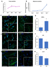
The functional characterization of recellularized rat livers. The amounts of (A) urea and (B) albumin secreted by the recellularized livers determined on days 1, 3, 5, and 7 were determined for the HM stage. Images of the immunohistochemistry analysis for (C) HNF4a, (E) AFP, and (G) Ki-67 in recellularized rat livers in the IHC and HM stages. Fluorescence intensity quantification of the immunohistochemistry images showing changes in the expression levels of (D) HNF4a, (F) AFP, and (H) Ki-67 in the IHC and HM stages. Three scaffolds at the IHC stage and three scaffolds in the IHC+ stages were analyzed for quantification. (* indicates statistically significant differences (p < 0.05); scale bars = 200 µm; scale bars for insets = 50 µm.)

References

    1. Byass P. The global burden of liver disease: A challenge for methods and for public health. BMC Med. 2014;12:159. doi: 10.1186/s12916-014-0159-5. - DOI - PMC - PubMed
    1. Starzl T.E., Marchioro T.L., Kaulla K.N.V., Hermann G., Brittain R.S., Waddell W.R. Homotransplantation of the liver in humans. Surg. Gynecol. Obstet. 1963;117:659–676. - PMC - PubMed
    1. United Network for Organ Sharing (UNOS) Transplant Trends. [(accessed on 4 May 2020)]. Available online: https://unos.org/data/transplant-trends/
    1. Badylak S.F., Taylor D., Uygun K. Whole-organ tissue engineering: Decellularization and recellularization of three-dimensional matrix scaffolds. Annu. Rev. Biomed. Eng. 2011;13:27–53. doi: 10.1146/annurev-bioeng-071910-124743. - DOI - PMC - PubMed
    1. Sabetkish S., Kajbafzadeh A.-M., Sabetkish N., Khorramirouz R., Akbarzadeh A., Seyedian S.L., Pasalar P., Orangian S., Beigi R.S.H., Aryan Z., et al. Whole-organ tissue engineering: Decellularization and recellularization of three-dimensional matrix liver scaffolds. J. Biomed. Mater. Res. A. 2015;103:1498–1508. doi: 10.1002/jbm.a.35291. - DOI - PubMed

LinkOut - more resources