

Decreased Reactive Oxygen Species Signaling Alters Glutamate Receptor Transport to Synapses in C. elegans AVA Neurons
- PMID: 35622512
- PMCID: PMC9007496
- DOI: 10.17912/micropub.biology.000528
Decreased Reactive Oxygen Species Signaling Alters Glutamate Receptor Transport to Synapses in C. elegans AVA Neurons
Abstract
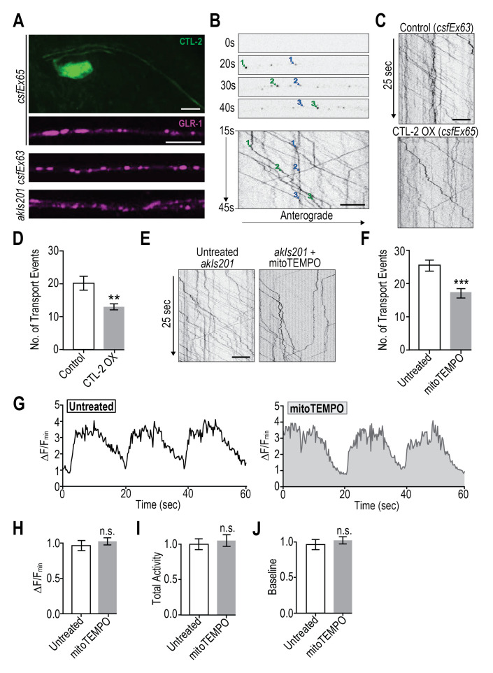
Reactive oxygen species (ROS) are chemically reactive molecules normally produced during cellular respiration. High ROS levels negatively impact forms of synaptic plasticity that rely on changes in the number of ionotropic glutamate receptors (iGluRs) at synapses. More recently, we have shown that physiological increases in ROS reduce iGluR transport to synapses by acting on activity-dependent calcium signaling. Here, we show that decreasing mitochondria-derived ROS decrease iGluR transport albeit in a calcium-independent manner. These data demonstrate differential regulatory mechanisms by elevated or diminished ROS levels which further support a physiological signaling role for ROS in regulating iGluR transport to synapses.
Figures
Similar articles
-
Neuron-specific regulation of associative learning and memory by MAGI-1 in C. elegans.PLoS One. 2009 Jun 24;4(6):e6019. doi: 10.1371/journal.pone.0006019. PLoS One. 2009. PMID: 19551147 Free PMC article.
-
The C. elegans nuclear receptor gene fax-1 and homeobox gene unc-42 coordinate interneuron identity by regulating the expression of glutamate receptor subunits and other neuron-specific genes.Dev Biol. 2005 Nov 1;287(1):74-85. doi: 10.1016/j.ydbio.2005.08.032. Epub 2005 Sep 23. Dev Biol. 2005. PMID: 16183052
-
The receptor protein tyrosine phosphatase CLR-1 is required for synaptic partner recognition.PLoS Genet. 2018 May 9;14(5):e1007312. doi: 10.1371/journal.pgen.1007312. eCollection 2018 May. PLoS Genet. 2018. PMID: 29742100 Free PMC article.
-
Ionotropic glutamate receptors in Caenorhabditis elegans.Neurosignals. 2003 May-Jun;12(3):108-25. doi: 10.1159/000072159. Neurosignals. 2003. PMID: 12904685 Review.
-
Activity-dependent regulation of vesicular glutamate and GABA transporters: a means to scale quantal size.Neurochem Int. 2006 May-Jun;48(6-7):643-9. doi: 10.1016/j.neuint.2005.12.029. Epub 2006 Mar 20. Neurochem Int. 2006. PMID: 16546297 Review.
Cited by
-
Activity-dependent mitochondrial ROS signaling regulates recruitment of glutamate receptors to synapses.Elife. 2024 Mar 14;13:e92376. doi: 10.7554/eLife.92376. Elife. 2024. PMID: 38483244 Free PMC article.
References
-
- Ashby, M., Daw, M., Issac, J., 2008. AMPA Receptors, in: Gereau, R., Swanson, G. (Eds.), The Glutamate Receptors. Humana Press, Totowa, NJ, pp. 1–44.
LinkOut - more resources
Full Text Sources
Research Materials