. 2022 Jun 14;17(6):1366-1379.
doi: 10.1016/j.stemcr.2022.05.001. Epub 2022 May 26.

Altered patterning of trisomy 21 interneuron progenitors

Affiliations

Altered patterning of trisomy 21 interneuron progenitors

Yathindar Giffin-Rao et al. Stem Cell Reports. .

Abstract

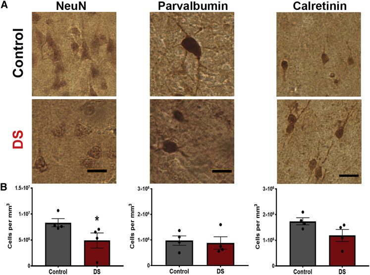
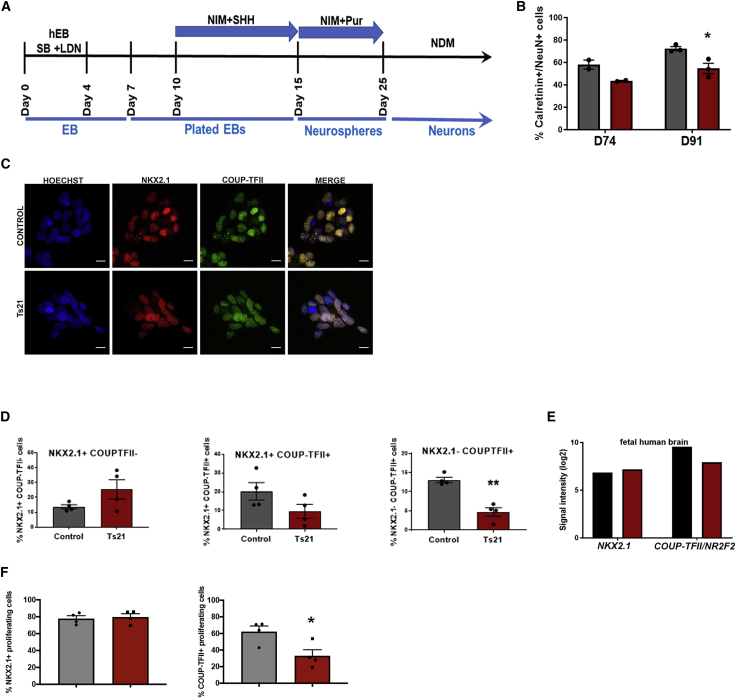
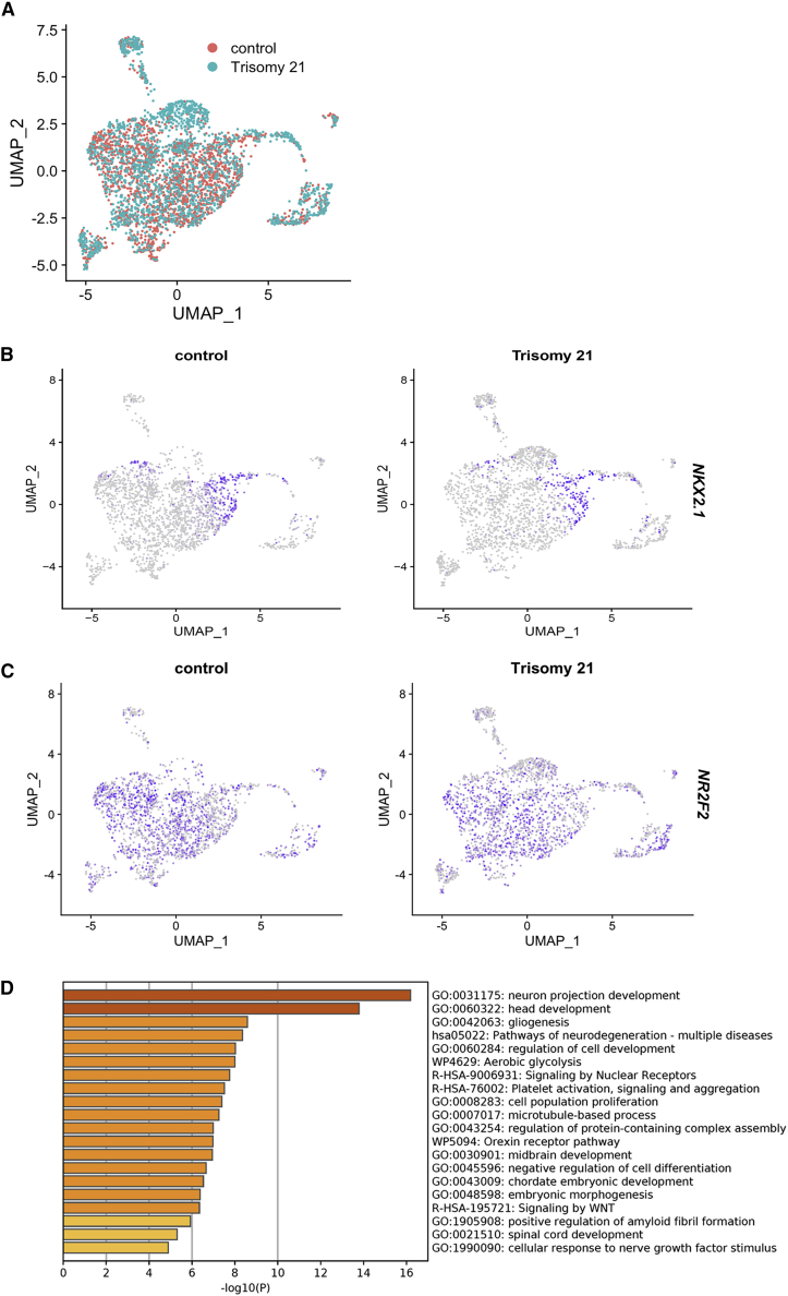
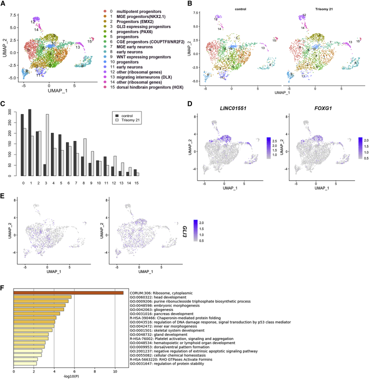
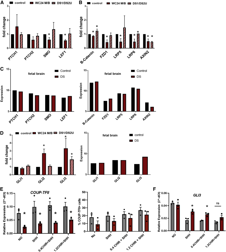
Individuals with Down syndrome (DS; Ts21), the most common genetic cause of intellectual disability, have smaller brains that reflect fewer neurons at pre- and post-natal stages, implicating impaired neurogenesis during development. Our stereological analysis of adult DS cortex indicates a reduction of calretinin-expressing interneurons. Using Ts21 human induced pluripotent stem cells (iPSCs) and isogenic controls, we find that Ts21 progenitors generate fewer COUP-TFII+ progenitors with reduced proliferation. Single-cell RNA sequencing of Ts21 progenitors confirms the altered specification of progenitor subpopulations and identifies reduced WNT signaling. Activation of WNT signaling partially restores the COUP-TFII+ progenitor population in Ts21, suggesting that altered WNT signaling contributes to the defective development of cortical interneurons in DS.

Keywords: Cortical development; Down syndrome; Neurogenesis; human; iPSCs; isogenic; neural differentiation; trisomy 21.


Figures

Figure 1
Figure 1
Reduced density of neurons in post-mortem Down syndrome cortex (A) Representative images of NeuN+, PV+, and CR+ neurons detected by immunohistochemistry (DAB) in control and DS post-mortem superior temporal gyrus (STG) samples. (B) Stereological quantification of the density of each neuron subtype in control and Down syndrome (DS) tissue. N = 4; NeuN p = 0.03, PV p = 0.89, CR p = 0.11 calculated using non-parametric Mann-Whitney U test.
Figure 2
Figure 2
Ts21 iPSCs generate fewer CR+ interneurons and fewer COUP-TFII+ progenitors (A) Interneuron progenitor differentiation protocol. (B) Proportion of calretinin (CR) neurons/(NeuN+) differentiated from control and Ts21 progenitors at days 74 and 91. p = 0.023 using unpaired t test with two-stage step up (Benjamini, Krieger, and Yekutieli).Control is black and Ts21 is red. (C) Immunofluorescence images of NKX2.1+ and COUP-TFII+ nuclei in control and Ts21 neural progenitor cells (NPCs). (D) Proportion of NKX2.1+ COUP-TFII- cells (MGE), NKX2.1+ COUP-TFII+ cells (caudal MGE), and NKX2.1- COUP-TFII+ cells (CGE) in control and Ts21 NPCs. ∗∗p < 0.001 using unpaired t test, N = 4 cell lines. (E) Expression of NKX2.1 and COUP-TFII in human fetal control and DS brain (14–17 weeks gestation, dorsolateral forebrain) (Olmos-Serrano et al., 2016). Control is black and Ts21 is red. (F) NKX2.1+/EdU+ and COUP-TFII+/EdU+ proliferating cells in the Ts21 cells compared with controls. p < 0.05 using unpaired t test, N = 4.
Figure 3
Figure 3
Single-cell RNA-seq reveals gene-expression differences in Ts21 progenitors (A) Dimensional reduction by experimental group (control and Ts21). (B) Feature plot of expression of NKX2.1 in control and trisomy cells. (C) Feature plot of expression of COUP-TFII/NR2F2 in control and trisomy cells. (D) Pathway analysis of differentially expressed genes in Ts21 compared with control.
Figure 4
Figure 4
Single-cell RNA-seq reveals differences in Ts21 progenitor clustering (A) Clustering analysis revealed 15 subpopulations of cells based on expression of known gene markers. (B) Clustering of control (2,134 cells) and Ts21 (2,158 cells). (C) Proportion of control and Ts21 cells in each cluster reveals enrichment of cluster 3 in Ts21 cells. (D) Feature plot of LINC0551 and FOXG1, markers that identify cluster 3 (Table S4). (E) Feature plot showing expression of GLI3. (F) Pathway analysis of differentially expressed genes in cluster 3 in Ts21 compared with control.
Figure 5
Figure 5
Ts21 ventral progenitors have decreased WNT and increased GLI3 expression (A and B) Quantitative PCR of SHH pathway genes (A) WNT signaling genes (B) in two iPSC lines of Ts21 and isogenic control lines. (C) Expression of SHH and WNT pathway genes in human fetal control and DS brain (14–17 weeks gestation, dorsolateral forebrain, Olmos-Serrano et al., 2016). (D) Quantitative PCR of GLI genes in progenitor cells and in human fetal control and DS brain (Olmos-Serrano et al., 2016). Statistical significance was determined by one-sample t test on ddCt values. p < 0.05, N = 3 for each gene/line. (E) The effects of activation of WNT via addition of a WNT agonist (CHIR, 0.4 and 1.2 μm) with SHH on COUP-TFII/NR2F2 gene expression, proportion of COUP-TFII+ cells, and GLI3 expression. Statistical significance was determined by one-sample t test on ddCt values. p < 0.05, N = 3 replicates in one pair of isogenic control and Ts21 cells.

References

    1. Adorno M., di Robilant B.N., Sikandar S.S., Acosta V.H., Antony J., Heller C.H., Clarke M.F. Usp16 modulates Wnt signaling in primary tissues through Cdkn2a regulation. Sci. Rep. 2018;8:17506. - PMC - PubMed
    1. Alfano C., Magrinelli E., Harb K., Studer M. The nuclear receptors COUP-TF: a long-lasting experience in forebrain assembly. Cell Mol. Life Sci. 2014;71:43–62. - PMC - PubMed
    1. Anderson S.A. Determination of cell fate within the telencephalon. Chem. senses. 2002;27:573–575. - PubMed
    1. Anderson S.A., Kaznowski C.E., Horn C., Rubenstein J.L.R., McConnell S.K. Distinct origins of neocortical projection neurons and interneurons in vivo. Cereb. Cortex. 2002;12:702–709. - PubMed
    1. Arshad A., Vose L.R., Vinukonda G., Hu F., Yoshikawa K., Csiszar A., Brumberg J.C., Ballabh P. Extended Production of cortical Interneurons into the third Trimester of human gestation. Cereb. Cortex. 2016;26:2242–2256. - PMC - PubMed

Publication types