. 2022 May 27;12(1):8923.
doi: 10.1038/s41598-022-12859-1.

Morphological characteristics of the infrapatellar fat pad

Affiliations

Morphological characteristics of the infrapatellar fat pad

Mutsuaki Edama et al. Sci Rep. .

Abstract

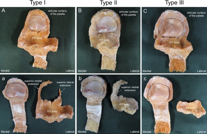
The relationship between the morphological characteristics of the infrapatellar fat pad (IFP) and joint deformity has yet to be fully elucidated. Therefore, the purpose of this study was to clarify the morphological characteristics of the IFP and to identify the relationships between morphological characteristics of the IFP and degenerative grade of the articular surface of the patella. This investigation examined 41 legs from 25 Japanese cadavers. The IFP length, width, and volume were measured. It was categorized into three types: Type I, IFP proximal located on medial and lateral sides of the patella; Type II, the IFP proximal only located medially; and Type III, absence of the IFP proximal. Articular surfaces were graded as macroscopically intact or mildly altered (Grade I), moderately (Grade II), or severely (Grade III). Grade III was significantly more frequent than Grades I or II in Type III. IFP volume was significantly larger in Type I than in Types II or III. A negative correlation was found between the degenerative grade of the articular surface of the patella and IFP volume. It was suggested that a relationship between the degenerative grade of the articular surface of the patella and the IFP volume.


Conflict of interest statement

The authors declare no competing interests.

Figures

Figure 1
Figure 1
The correlation of between degenerative grade of the articular surface of the patella and IFP volume. Grade I: macroscopically intact or only mildly altered; Grade II: macroscopically moderately altered (if fissuring or fibrillation is observed); Grade III: macroscopically severely altered (if eburnation is present).
Figure 2
Figure 2
Method of dissection; right knee. (A) Medial view. The synovium and medial meniscus are incised along the anterior edge of the medial collateral ligament, and the synovium and meniscus are lifted forward. (B) Lateral view. The synovium and lateral meniscus are incised along the anterior edge of the lateral collateral ligament, and the synovium and meniscus are lifted forward. (C) Anterior view. Knee flexion position. The quadriceps femoris and patella are inverted from proximal to distal. White arrow: ligamentum mucosum.
Figure 3
Figure 3
Method for dissection of the infrapatellar fat pad; right knee. (A) Medial view. The vastus medialis muscle is flipped forward and the synovium is gripped with forceps. An incision is made along the white dashed line to remove synovium not adherent to the infrapatellar fat pad. (B) Posterior view. The quadriceps femoris is removed along the morphology of the infrapatellar fat. IFP infrapatellar fat pad, SFP suprapatellar fat pad. White dashed line: boundary between Infrapatellar fat pad and synovitis.
Figure 4
Figure 4
Measurement of the infrapatellar fat pad; right knee, posterior view. (A) Length of the infrapatellar fat pad superior medial extension and superior lateral extension, infrapatellar fat pad body. (B) Width of the infrapatellar fat pad superior medial extension and superior lateral extension, infrapatellar fat pad body. Black arrow 1: In cases with the infrapatellar fat pad superior medial extension connecting to the suprapatellar fat pad. Black arrow 2: In cases with the infrapatellar fat pad superior lateral extension not connecting to the suprapatellar fat pad. Black arrow 3: Infrapatellar fat pad body. Black arrow 4: Infrapatellar fat superior medial extension top. Black arrow 5: Infrapatellar fat superior medial extension center. Black arrow 6: Infrapatellar fat superior medial extension end. Black arrow 7: Infrapatellar fat superior lateral extension top. Black arrow 8: Infrapatellar fat superior lateral extension center. Black arrow 9: Infrapatellar fat superior lateral extension end. Black arrow 10: Infrapatellar fat pad body. White line: Reference line.
Figure 5
Figure 5
Classification of the IFP. (A) Type I: IFP proximal located on the medial side (superior medial extension) and lateral side (superior lateral extension) of the patella. (B) Type II: only IFP proximal located on the medial side (superior medial extension) of the patella. (C) Type III: absence of IFP proximal located on the medial and lateral sides of the patella. (ac) IFP removed from the patella. IFP infrapatellar fat pad.
Figure 6
Figure 6
Classification of degenerative grade of the articular surface. (A) Grade I: macroscopically intact or only mildly altered. (B) Grade II: macroscopically moderately altered (if fissuring or fibrillation is observed). (C) Grade III: macroscopically severely altered (if eburnation is present).

References

    1. Hoffa A. The influence of the adipose tissue with regard to the pathology of the knee joint. J. Am. Med. Assoc. 1904;XLIII:795–796. doi: 10.1001/jama.1904.92500120002h. - DOI
    1. Mac CM. The movements of bones and joints; The synovial fluid and its assistants. J. Bone Jt. Surg. Br. 1950;32-b:244–252. doi: 10.1302/0301-620x.32b2.244. - DOI - PubMed
    1. Duri ZA, Aichroth PM, Dowd G. The fat pad. Clinical observations. Am. J. Knee Surg. 1996;9:55–66. - PubMed
    1. Biedert RM, Sanchis-Alfonso V. Sources of anterior knee pain. Clin. Sports Med. 2002;21:335–347. doi: 10.1016/s0278-5919(02)00026-1. - DOI - PubMed
    1. Gallagher J, Tierney P, Murray P, O'Brien M. The infrapatellar fat pad: Anatomy and clinical correlations. Knee Surg. Sports Traumatol. Arthrosc. 2005;13:268–272. doi: 10.1007/s00167-004-0592-7. - DOI - PubMed

Publication types