. 2022 Dec;29(12):2347-2361.
doi: 10.1038/s41418-022-01021-z. Epub 2022 May 27.

A truncating variant of RAD51B associated with primary ovarian insufficiency provides insights into its meiotic and somatic functions

Affiliations

A truncating variant of RAD51B associated with primary ovarian insufficiency provides insights into its meiotic and somatic functions

Monica M Franca et al. Cell Death Differ. 2022 Dec.

Abstract

Primary ovarian insufficiency (POI) causes female infertility by abolishing normal ovarian function. Although its genetic etiology has been extensively investigated, most POI cases remain unexplained. Using whole-exome sequencing, we identified a homozygous variant in RAD51B -(c.92delT) in two sisters with POI. In vitro studies revealed that this variant leads to translation reinitiation at methionine 64. Here, we show that this is a pathogenic hypomorphic variant in a mouse model. Rad51bc.92delT/c.92delT mice exhibited meiotic DNA repair defects due to RAD51 and HSF2BP/BMRE1 accumulation in the chromosome axes leading to a reduction in the number of crossovers. Interestingly, the interaction of RAD51B-c.92delT with RAD51C and with its newly identified interactors RAD51 and HELQ was abrogated or diminished. Repair of mitomycin-C-induced chromosomal aberrations was impaired in RAD51B/Rad51b-c.92delT human and mouse somatic cells in vitro and in explanted mouse bone marrow cells. Accordingly, Rad51b-c.92delT variant reduced replication fork progression of patient-derived lymphoblastoid cell lines and pluripotent reprogramming efficiency of primary mouse embryonic fibroblasts. Finally, Rad51bc.92delT/c.92delT mice displayed increased incidence of pituitary gland hyperplasia. These results provide new mechanistic insights into the role of RAD51B not only in meiosis but in the maintenance of somatic genome stability.


Conflict of interest statement

The authors declare no competing interests.

Figures
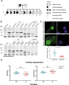
Fig. 1
Fig. 1. The variant c.92delT leads to translation re-initiation at AUG 64 and to altered nuclear localization.
A Family Pedigree. A homozygous recessive variant in RAD51B was shown to be present in two sisters from Brazil affected with POI. The black arrow indicates proband (II-1). Pedigree numbers of individuals are indicated below the symbols. Samples sent for whole-exome sequencing (WES) are indicated by a hash and samples sent for Sanger sequencing are indicated by an asterisk. Sanger electropherograms confirmed the presence of the homozygous variant in both affected sisters II-1 and II-4. B HEK293T cells were transfected or not (Vehicle) with the different variants of RAD51B: the WT form (WT), the c.92delT form and both the WT and c.92delT forms in which the three secondary Methionines were mutated into Alanines (individual Met to Ala substituting the M39 to A39, the M55 to A55 and the M64 to A64; double Met to Ala substitutions following the above argument, both M39A39 and M55A55, both M39A39 and M64A64 and both M55A55 and M64A64. Of note, the bigger size of the RAD51B products is due to the presence of GFP tag (27 kDa). C COS7 cells were transfected to express human WT or mutant RAD51B fused to GFP tag. The WT construct showed a robust nuclear signal in addition to a faint cytoplasmic pattern. In contrast, the mutant variant displayed a partial delocalization of the nuclear signal to the cytoplasm. Quantification of the nucleus/cytoplasm signal rate is shown in the lower plot. Scale bars: 20 µm. D Fertility assessment in female mice of WT and mutant Rad51b. The plots show the number of pups per litter and the litters per month. Rad51bc.92delT/c.92delT variant is referred as Rad51bKI/KI for simplicity. Welch´s t-test analysis: ns, non-significant differences; ****p < 0.0001.
Fig. 2
Fig. 2. Rad51bKI/KI mice show defects in DNA repair.
A Double immunolabelling of γH2AX (green) and SYCP3 (red) of spermatocyte and oocyte spreads from WT and KI mice showing the accumulation of γH2AX patches in the mutant pachynemas. Plots below the panels show the quantification of γH2AX intensity. B Double immunolabelling of RAD51 (green) and SYCP3 (red) of spermatocyte and oocyte spreads from Rad51WT/WT and Rad51bKI/KI. RAD51 foci accumulate at pachytene in KI spermatocytes and at diplotene in KI oocytes (small green dots). Plots below the panels represents the quantification of RAD51 foci on each genotype. Rad51bc.92delT/c.92delT variant is referred to as Rad51bKI/KI for simplicity. Welch´s t-test analysis: ns, non-significant differences; **p < 0.01; ***p < 0.001; ****p < 0.0001. Bar in panels, 10 μm. See Supplementary Table 5 for raw data quantification.
Fig. 3
Fig. 3. Rad51b mutant mice show an accumulation of BRME1 and HSF2BP and an abnormal CO formation.
A Double labelling of BRME1 (green) and SYCP3 (red) of spermatocyte and oocyte spreads from WT and KI mice showing the accumulation of BRME1 in mutant late pachynemas and diplonemas. Double labelling of HSF2BP (green) and SYCP3 (red) of meiocyte spreads from Rad51bWT/WT and Rad51bKI/KI mice showing the accumulation of HSF2BP in mutant late pachynemas and diplonemas. Plots on the right side of the panels represent the quantification of BRME1 and HSF2BP foci and intensity. B Double immunolabelling of MLH1 (green) and SYCP3 (red) of spermatocyte and oocyte spreads from Rad51bWT/WT and Rad51bKI/KI. MLH1 foci are significant reduced in mutant Rad51b meiocytes. The plots on the right of the panels represent the quantification of MLH1 foci at pachytene in both male and female meiocytes. Quantification of the % of spermatocytes with any autosome or the sexual bivalent without MLH1 foci. n = 108–114 (autosomes), n = 66–96 (X-Y bivalent). Quantification of the % of oocytes with any autosome without MLH1 foci. n = 51–54 (autosomes). Rad51bc.92delT/c.92delT variant is referred as Rad51bKI/KI for simplicity. Welch´s t-test analysis: ns, non-significant differences; *p < 0.05; **p < 0.01; ***p < 0.001; ****p < 0.0001. Bar in panels, 10 μm. See Supplementary Table 5 for raw data quantification.
Fig. 4
Fig. 4. Loss of interaction between RAD51B-c.92delT and RAD51C as well as a reduction of the interaction between RAD51B-c.92delT and HR-specific interactors as revealed by co-immunoprecipitation.
HEK293T cells were co-transfected with either RAD51B-WT or RAD51B-c.92delT and A RAD51C-HA, B Flag-RAD51, C HA-HELQ or D Flag-DMC1. Protein complexes were immunoprecipitated with either an anti-RAD51B, anti-Flag, anti-HA or IgGs (negative control), and analyzed by immunoblotting with the indicated antibody. The red > indicates the band corresponding to RAD51B.
Fig. 5
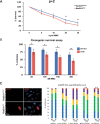
Fig. 5. Rad51b mutant MEFs show an increased susceptibility to MMC-induced DNA damage.
A Cell proliferation assay of WT and mutant Rad51b primary MEFs at passage 2 (p + 2) incubated in presence of a continuous treatment with MMC. The results are expressed as a percentage relative to the control (not treated with MMC). Each point on the graph represents the mean ± SD. B Percentage of colonies obtained by clonogenic cell survival assays after treatment with MMC. The results are expressed as a percentage relative to the control (untreated) of Rad51bWT/WT and Rad51bKI/KI immortalized cells. C Representative γH2AX immunolabelling of WT and mutant Rad51b at 72 h. Quantification of γH2AX foci in Rad51b WT and mutant MEFs. Rad51bWT/WT and Rad51bKI/KI MEFs at p + 2 were incubated in presence of MMC at 1 µg/ml for 1 h and then supplemented with fresh medium without MMC. The quantification was performed at different time points: 0 h: 0 h, 6 h: 6 h, 24 h: 24 h, 48 h; 48 h and 72 h: 72 h. Cells were classified in 5 groups: 0 foci, 1 to 10 foci, 10 to 30 foci, 30 to 150 foci and >150 foci. n = 3. Rad51bc.92delT/c.92delT variant is referred as Rad51bKI/KI for simplicity. Welch´s t-test analysis: *p < 0.05; **p < 0.01. Bar in panel, 10 µm.
Fig. 6
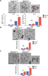
Fig. 6. MMC-induced CIN in mouse and human RAD51B-c.92delT cells.
A Evaluation of metaphase chromosome breaks/gaps from Rad51WT/WT and Rad51bKI/KI MEFs after MMC treatment (150 nM). Lower panel (graphs) shows the quantification of breaks/gaps at passage 2 (p + 2) and passage 4 (p + 4). B Evaluation of metaphase chromosome aberrations from bone marrow of Rad51bWT/WT and Rad51bKI/KI after intraperitoneal injection of MMC (4 mg/kg). In addition, to breaks/gaps, triradial chromosomes were observed only in the mutant mice (shown by red asterisks). C Homozygous RAD51B-c.92delT human-derived lymphoblastoid cells showed more chromosome alterations with and without MMC treatment (200 nM) in comparison with the corresponding heterozygous sister. n = 3. Rad51bc.92delT/c.92delT variant is referred to as Rad51bKI/KI for simplicity. Welch´s t-test analysis: ns, non-significant differences; *p < 0.5; **p < 0.01; ***p < 0.001. Bar in panels, 10 μm.
Fig. 7
Fig. 7. RAD51B-c.92delT have a role in replication fork homeostasis but not in SCE.
A Cell proliferation assay of WT and mutant Rad51b primary MEFs at passage 2 (p + 2) and 3 (p + 3) incubated in presence of a continuous treatment with hydroxyurea (HU) and in presence of a continuous treatment with Aphidicolin. The results are expressed as a percentage relative to the control (not treated). Each point on the graph represents the mean ± SD. B Top: Schematic of the stretched DNA fiber assay. Bottom: Examples of DNA fiber images from the indicated cells. Bar in panels, 10 µM. Histogram shows the fork progression rate (median and distribution) in each experimental condition. n = 3 experimental replicates (data pooled together). >400 structures scored per condition and replica. ***p < 0.001; in Mann-Whitney test. C Sister chromatid exchange (SCE) per chromosome after treatment with MMC. RAD51BWT/c.92delT and RAD51Bc.92delT/c.92delT variants are referred to as RAD51BWT/KI and RAD51BKI/KI for simplicity. Welch´s t-test analysis: ns, non-significant differences, *p < 0.5; **p < 0.01.
Fig. 8
Fig. 8. RAD51B-c.92delT leads to a reduced reprogramming efficiency of MEFs and humanized Rad51bKI/KI mice show an increased incidence of hyperplasia of the pituitary gland.
A MEFs from the indicated genotypes were infected with the 3 reprogramming factors and the numbers of alkaline phosphatase positive colonies were counted showing a significantly reduction (up to ~2 fold) in Rad51b-c.92delT homozygous mutant MEFs in comparison with the wild-type control. n = 9. Welch´s t-test analysis: ***p < 0.001. B Macroscopic image of adenohypophysis from Rad51bWT/WT and adenohypophysis rom Rad51bKI/KI mice. Reticulin staining (CH) and Prolactin IHQ (I, J) of pituitary adenohypophysis from Rad51bWT/WT and Rad51bKI/KI. C, D Normal adenohypophysis from Rad51bWT/WT show a reticulin staining pattern that is partially lost in zones of hyperplasia where the cell size is increased (magnified in D, indicated by an arrow). E, F Microadenoma from a Rad51bKI/KI showing a complete loss of reticulin staining pattern that is still persistent in the neighbor misplaced adenohypophysis (magnified in F). G, H Macroadenoma from a Rad51bKI/KI showing total loss of reticulin staining pattern and complete absence of normal adenohypophysis tissue. The neoplasm shows different adenoid and pseudopapillary growing patterns (magnified in H). I, J IHQ of prolactin in normal pituitary glands from Rad51bWT/WT showing labelled cells unevenly distributed whereas adenomas from Rad51bKI/KI revealed a high density of prolactin expressing cells. Bar in panels, 250 μm (C, E, G) and 50 μm (D, F, H, I, J). Rad51bc.92delT/c.92delT variant is referred as Rad51bKI/KI for simplicity.

References

    1. Handel MA, Schimenti JC. Genetics of mammalian meiosis: regulation, dynamics and impact on fertility. Nat Rev Genet. 2010;11:124–36. doi: 10.1038/nrg2723. - DOI - PubMed
    1. Paiano J, Wu W, Yamada S, Sciascia N, Callen E, Paola Cotrim A, et al. ATM and PRDM9 regulate SPO11-bound recombination intermediates during meiosis. Nat Commun. 2020;11:857. doi: 10.1038/s41467-020-14654-w. - DOI - PMC - PubMed
    1. Zhao W, Vaithiyalingam S, San Filippo J, Maranon DG, Jimenez-Sainz J, Fontenay GV, et al. Promotion of BRCA2-Dependent Homologous Recombination by DSS1 via RPA Targeting and DNA Mimicry. Mol Cell. 2015;59:176–87. doi: 10.1016/j.molcel.2015.05.032. - DOI - PMC - PubMed
    1. Martinez JS, von Nicolai C, Kim T, Ehlen A, Mazin AV, Kowalczykowski SC, et al. BRCA2 regulates DMC1-mediated recombination through the BRC repeats. Proc Natl Acad Sci USA. 2016;113:3515–20. doi: 10.1073/pnas.1601691113. - DOI - PMC - PubMed
    1. Chun J, Buechelmaier ES, Powell SN. Rad51 paralog complexes BCDX2 and CX3 act at different stages in the BRCA1-BRCA2-dependent homologous recombination pathway. Mol Cell Biol. 2013;33:387–95. doi: 10.1128/MCB.00465-12. - DOI - PMC - PubMed

Publication types