. 2022 Apr 22;11(5):819.
doi: 10.3390/antiox11050819.

Metabolic Profiling, Chemical Composition, Antioxidant Capacity, and In Vivo Hepato- and Nephroprotective Effects of Sonchus cornutus in Mice Exposed to Cisplatin

Affiliations

Metabolic Profiling, Chemical Composition, Antioxidant Capacity, and In Vivo Hepato- and Nephroprotective Effects of Sonchus cornutus in Mice Exposed to Cisplatin

Sameh S Elhady et al. Antioxidants (Basel). .

Abstract

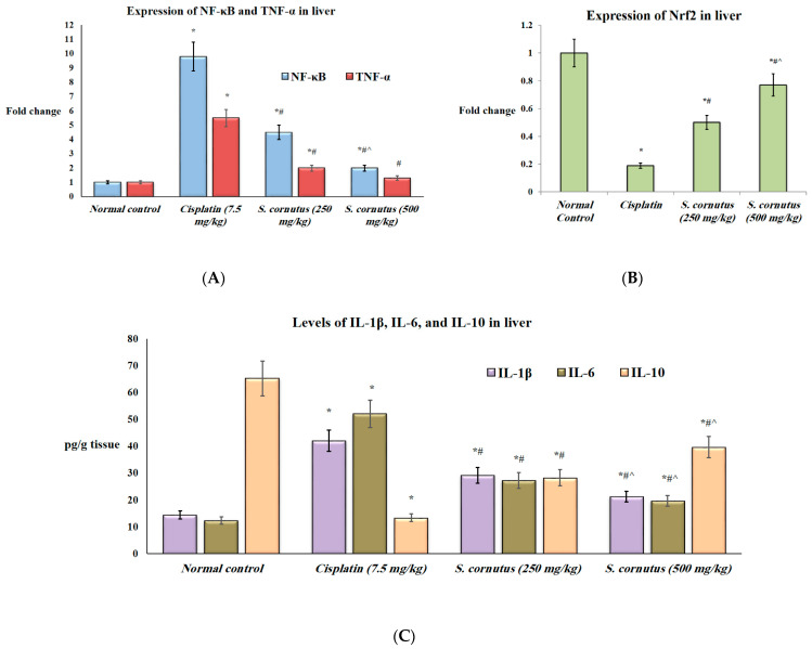
Sonchus cornutus (Asteraceae) is a wild. edible plant that represents a plentiful source of polyphenolic compounds. For the first time, the metabolic analysis profiling demonstrated the presence of anthocyanidin glycosides, coumarins, flavonoids and their corresponding glycosides, and phenolic acids. The total phenolic compounds were determined to be 206.28 ± 14.64 mg gallic acid equivalent/gm, while flavonoids were determined to be 45.56 ± 1.78 mg quercetin equivalent/gm. The crude extract of S. cornutus exhibited a significant 1,1-diphenyl-2-picrylhydrazyl free radical scavenging effect with half-maximal inhibitory concentration (IC50) of 16.10 ± 2.14 µg/mL compared to ascorbic acid as a standard (10.64 ± 0.82 µg/mL). In vitro total antioxidant capacity and ferric reducing power capacity assays revealed a promising reducing potential of S. cornutus extract. Therefore, the possible protective effects of S. cornutus against hepatic and renal toxicity induced by cisplatin in experimental mice were investigated. S. cornutus significantly ameliorated the cisplatin-induced disturbances in liver and kidney functions and oxidative stress, decreased MDA, ROS, and NO levels, and restored CAT and SOD activities. Besides, it reversed cisplatin-driven upregulation in inflammatory markers, including iNOS, IL-6, and IL-1β levels and NF-κB and TNF-α expression, and elevated anti-inflammatory IL-10 levels and Nrf2 expression. Additionally, the extract mitigated cisplatin alteration in apoptotic (Bax and caspase-3) and anti-apoptotic (Bcl-2) proteins. Interestingly, hepatic, and renal histopathology revealed the protective impacts of S. cornutus against cisplatin-induced pathological changes. Our findings guarantee a protective effect of S. cornutus against cisplatin-induced hepatic and renal damage via modulating oxidative stress, inflammation, and apoptotic pathways.

Keywords: Sonchus cornutus; apoptosis; drug discovery; hepatotoxicity; industries development; inflammation; nephrotoxicity; oxidative stress; polyphenolic compounds.


Conflict of interest statement

The authors declare no conflict of interest.

Figures

Figure 1
Figure 1
Structures of the detected metabolites as listed in Table 1.
Figure 2
Figure 2
The effect of S. cornutus crude extract on the expression and protein levels of inflammtory markers in the liver tissue of the experimental mice. (A) NF-κβ and TNF-α expression, (B) Nrf2 expression, (C) Il-1β, IL-6, and IL-10 levels. NF-κβ = nuclear factor kappa B; TNF-α = tumor necrosis factor-α; Nrf2 = nuclear factor-erythroid factor 2-related factor 2; IL = interleukin. Data are expressed as mean ± SD and analyzed using one-way ANOVA followed by Bonferroni’s post hoc test (n = 8). * significantly different vs. the normal control group; # significantly different vs. the cisplatin group; ^ significantly different vs. the S. cornutus (250 mg/kg) group. Differences were considered significant at p < 0.05.
Figure 3
Figure 3
The effect of S. cornutus crude extract on the expression and protein levels of inflammtory markers in the kidney tissue of the experimental mice. (A) NF-κβ and TNF-α expression, (B) Nrf2 expression, (C) Il-1β, IL-6, and IL-10 levels. NF-κβ = nuclear factor kappa B; TNF-α = tumor necrosis factor-α; Nrf2 = nuclear factor-erythroid factor 2-related factor 2; IL = interleukin. Data are expressed as mean ± SD and analyzed using one-way ANOVA followed by Bonferroni’s post hoc test (n = 8). * significantly different vs. the normal control group; # significantly different vs. the cisplatin group; ^ significantly different vs. the S. cornutus (250 mg/kg) group. Differences were considered significant at p < 0.05.
Figure 4
Figure 4
The effect of S. cornutus crude extract on the expression levels of caspase-3 determined by immunohistochemistry in the (A) liver and (B) kidney tissues of the experimental mice. Data of the percentage of positive stained area are expressed as mean ± SD and analyzed using one-way ANOVA followed by Bonferroni’s post hoc test (n = 8). * significantly different vs. the normal control group; # significantly different vs. the cisplatin group; ^ significantly different vs. the S. cornutus (250 mg/kg) group. Differences were considered significant at p < 0.05.
Figure 5
Figure 5
The effect of S. cornutus crude extract on the expression levels of Bax determined by immunohistochemistry in the (A) liver and (B) kidney tissues of the experimental mice. Data of the percentage of positive stained area are expressed as mean ± SD and analyzed using one-way ANOVA followed by Bonferroni’s post hoc test (n = 8). * significantly different vs. the normal control group; # significantly different vs. the cisplatin group; ^ significantly different vs. the S. cornutus (250 mg/kg) group. Differences were considered significant at p < 0.05.
Figure 6
Figure 6
The effect of S. cornutus crude extract on the expression levels of Bcl-2 determined by immunohistochemistry in the (A) liver and (B) kidney tissues of the experimental mice. Data of the percentage of positive stained area are expressed as mean ± SD and analyzed using one-way ANOVA followed by Bonferroni’s post hoc test (n = 8). * significantly different vs. the normal control group; # significantly different vs. the cisplatin group; ^ significantly different vs. the S. cornutus (250 mg/kg) group. Differences were considered significant at p < 0.05.
Figure 7
Figure 7
A photomicrography of liver tissue (H&E, ×200). (A) normal control group; shows normal hepatic architecture, with clear central vein (CV) and hepatocytes radially arranged in plate (black arrows), and normal sinusoidal spaces between them (red arrows). (B) cisplatin group; shows infiltration of inflammatory cells (black arrows), with congested central vein (CV) and dilated sinusoidal spaces (red arrows). (C) S. cornutus (250 mg/kg) group; shows partial improvement, less infiltration of inflammatory cells (black arrows), but still dilated sinusoidal spaces (red arrows). (D) S. cornutus (500 mg/kg) group; shows the best improvement, nearly normal hepatic architecture, and restoration of normal sinusoidal spaces (red arrows).
Figure 8
Figure 8
A photomicrography of kidney tissue (H&E, ×100). (A) normal control group; shows normal histology, with normal looking glomeruli (asterisks), proximal tubules (black arrows), and distal tubules (blue arrows). (B) cisplatin group; shows patchy interstitial inflammatory cell infiltrate (black arrows), congestion (blue arrows), focal tubular cell necrosis with condensed eosinophilic cytoplasm (green arrows), and regenerative changes with slightly basophilic cytoplasm and enlarged nuclei (double headed arrows). (C) S. cornutus (250 mg/kg) group; shows patchy interstitial inflammatory cell infiltrate (black arrows), congestion (blue arrows), focal tubular cell necrosis with condensed eosinophilic cytoplasm (green arrows), and regenerative changes with slightly basophilic cytoplasm and enlarged nuclei (double headed arrows). (D) S. cornutus (500 mg/kg) group; shows patchy interstitial inflammatory cell infiltrate (black arrows), mild congestion (blue arrows), minimal focal tubular cell necrosis (green arrows), and some regenerative changes with slightly basophilic cytoplasm and enlarged nuclei (double headed arrows).

References

    1. Ghosh S. Cisplatin: The first metal based anticancer drug. Bioorg. Chem. 2019;88:102925. doi: 10.1016/j.bioorg.2019.102925. - DOI - PubMed
    1. Singh N., Magotra R., Sharma A.K., Ahmed M., Khajuria V. Effect of cisplatin on liver of male albino rats. J. Evol. Med. Dent. Sci. 2015;4:8993–8998. doi: 10.14260/jemds/2015/1305. - DOI
    1. Abdel Moneim A.E., Othman M.S., Aref A.M. Azadirachta indica attenuates cisplatin-induced nephrotoxicity and oxidative stress. Biomed. Res. Int. 2014;2014:647131. doi: 10.1155/2014/647131. - DOI - PMC - PubMed
    1. Ma L., Wang H., Wang C., Su J., Xie Q., Xu L., Yu Y., Liu S., Li S., Xu Y. Failure of elevating calcium induces oxidative stress tolerance and imparts cisplatin resistance in ovarian cancer cells. Aging Dis. 2016;7:254–266. doi: 10.14336/AD.2016.0118. - DOI - PMC - PubMed
    1. Abouzed T., Soliman M., Khatab S., Elgazzar A., Gouda W., Dorghamm D. The protective impacts of Spriulina platensis against cisplatin-induced renal injury through the regulation of oxidative stress, pro-inflammatory cytokines and Bax/Bcl2 expression cascade. Toxicol. Res. 2022;11:169–178. doi: 10.1093/toxres/tfab128. - DOI - PMC - PubMed