. 2022 Apr 30;11(5):899.
doi: 10.3390/antiox11050899.

Thioredoxin-1 Ameliorates Oxygen-Induced Retinopathy in Newborn Mice through Modulation of Proinflammatory and Angiogenic Factors

Affiliations

Thioredoxin-1 Ameliorates Oxygen-Induced Retinopathy in Newborn Mice through Modulation of Proinflammatory and Angiogenic Factors

Junichi Ozawa et al. Antioxidants (Basel). .

Abstract

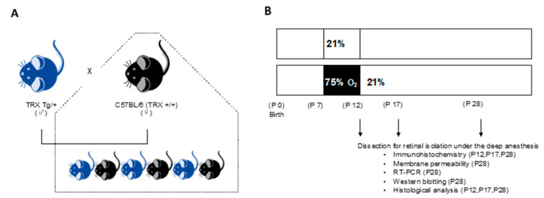
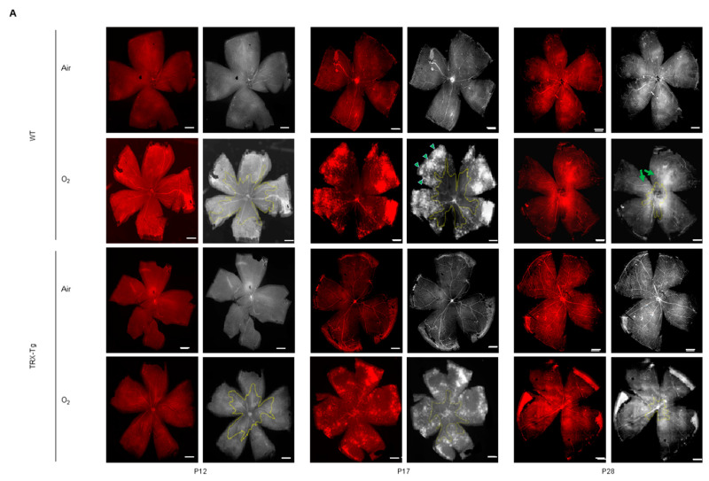
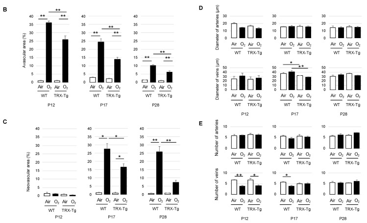
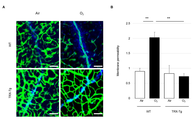
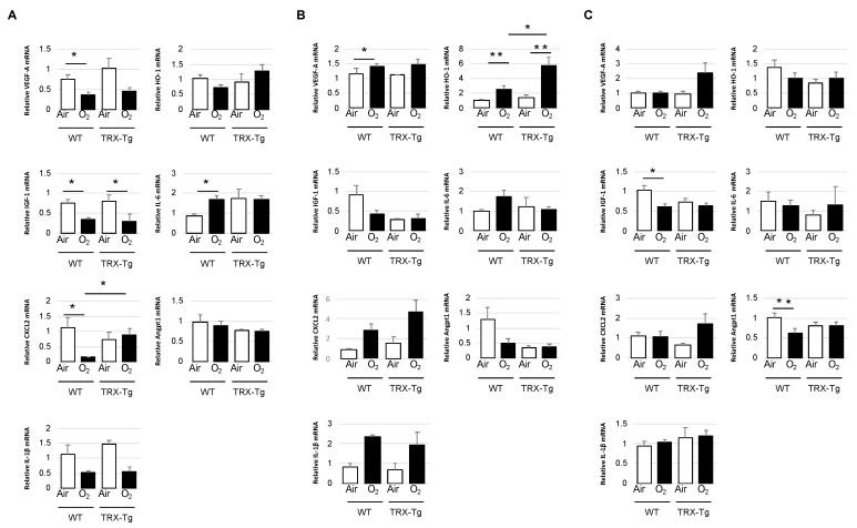
Oxygen-induced retinopathy (OIR) is an animal model for retinopathy of prematurity, which is a leading cause of blindness in children. Thioredoxin-1 (TRX) is a small redox protein that has cytoprotective and anti-inflammatory properties in response to oxidative stress. The purpose of this study was to determine the effect of TRX on OIR in newborn mice. From postnatal day 7, C57BL/6 wild type (WT) and TRX transgenic (TRX-Tg) mice were exposed to either 21% or 75% oxygen for 5 days. Avascular and neovascular regions of the retinas were investigated using fluorescence immunostaining. Fluorescein isothiocyanate-dextran and Hoechst staining were used to measure retinal vascular leakage. mRNA expression levels of proinflammatory and angiogenic factors were analyzed using quantitative polymerase chain reaction. Retinal histological changes were detected using immunohistochemistry. In room air, the WT mice developed well-organized retinas. In contrast, exposing WT newborn mice to hyperoxia hampered retinal development, increasing the retinal avascular and neovascular areas. After hyperoxia exposure, TRX-Tg mice had enhanced retinal avascularization compared with WT mice. TRX-Tg mice had lower retinal neovascularization and retinal permeability during recovery from hyperoxia compared with WT mice. In the early stages after hyperoxia exposure, VEGF-A and CXCL-2 expression levels decreased, while IL-6 expression levels increased in WT newborn mice. Conversely, no differences in gene expressions were observed in the TRX-Tg mouse retina. IGF-1 and Angpt1 levels did not decrease during recovery from hyperoxia in TRX-Tg newborn mice. As a result, overexpression of TRX improves OIR in newborn mice by modulating proinflammatory and angiogenic factors.

Keywords: angiogenic factor; hyperoxia; newborn mouse; oxygen-induced retinopathy; retinal blood vessel; retinopathy of prematurity; thioredoxin-1.


Conflict of interest statement

The authors have no affiliations with or involvement in any organization or entity with any financial or nonfinancial interest in the subject matter or materials discussed in this manuscript.

Figures

Figure 1
Figure 1
Experimental design. (A) The breeding scheme and genotypes of the mice that were studied. Heterozygous TRX-Tg male and WT female mice were mated to produce either TRX heterozygous or WT littermates. (B) Experimental method of exposing newborn mice to hyperoxia. P7 pups were randomly assigned to normal room air (21% O2) or hyperoxic (75% O2) conditions with a nursing mother. On P12, some mice were released to room air and were able to recover until P12, P17, and P28. TRX, thioredoxin-1; Tg, transgenic; WT, wild type; P, postnatal day.
Figure 2
Figure 2
Immunohistochemical staining of mouse retinal vessels with anti-CD31 antibody. (A) In each group, the representative original image (left column) and the processed image for analysis (right column). An avascular field is indicated by the yellow line. The green arrowheads indicate the neovascular area. Green arrows show tortuous blood vessels. Each scale bar is 250 μm. (B) around the optic disc, there is an avascular retinal region. (C) The neovascular zone. (D) The number of retinal arteries. (E) The number of retinal veins. * p < 0.05, ** p < 0.01, P12, n = 5; P17, n = 6; P28, n = 5; P, postnatal day; WT, wild type; TRX-Tg, thioredoxin-1 transgenic.
Figure 2
Figure 2
Immunohistochemical staining of mouse retinal vessels with anti-CD31 antibody. (A) In each group, the representative original image (left column) and the processed image for analysis (right column). An avascular field is indicated by the yellow line. The green arrowheads indicate the neovascular area. Green arrows show tortuous blood vessels. Each scale bar is 250 μm. (B) around the optic disc, there is an avascular retinal region. (C) The neovascular zone. (D) The number of retinal arteries. (E) The number of retinal veins. * p < 0.05, ** p < 0.01, P12, n = 5; P17, n = 6; P28, n = 5; P, postnatal day; WT, wild type; TRX-Tg, thioredoxin-1 transgenic.
Figure 3
Figure 3
Mouse retinal vascular permeability. (A) Original picture immunostaining for blood vessel permeability on P28. The green network depicts FITC-dextran stained blood vessels, whereas the blue network reveals Hoechst 3324-stained cells. The scale bar is 20 μm. (B) Blood vessel permeability is calculated by [(Hoechst 33342 area) − (FITC-dextran + area around the blood vessel stained with Hoechst 33342)]/(FITC-dextran area) in each group. ** p < 0.01. Results are displayed as mean ± standard error. WT room air, n = 4; WT hyperoxia, n = 6; TRX-Tg room air, n = 6; TRX-Tg hyperoxia, n = 6. WT, wild type; TRX-Tg, thioredoxin-1 transgenic.
Figure 4
Figure 4
Expression levels of tight junction protein in the mouse retina. (A) Occludin (60–82 kDa) was tested. β-actin (42 kDa) was used as a control. (B) Claudin-5 (23 kDa) was examined. β-actin was used as a control. The results are displayed as mean ± standard error. Six retinas from each group were used (n = 6). WT, wild type; TRX-Tg, thioredoxin-1 transgenic.
Figure 5
Figure 5
Mouse retinal mRNA expression levels of VEGF-A, IGF-1, CXCL2, IL-1β, HO-1, IL-6, and Angpt1. Quantitative real-time PCR was carried out at (A) P12, (B) P17, and (C) P28. * p < 0.05, ** p < 0.01. Results are displayed as mean ± standard error (n = 5–6). P, postnatal day; WT, wild type; TRX-Tg, thioredoxin-1 transgenic; VEGF-A, vascular endothelial growth factor-a; IGF-1, insulin-like growth factor-1; CXCL-2, chemokine ligand 2; IL-1β, interleukin-1β; HO-1, heme oxygenase-1; Il-6, interleukin-6; Angpt1, angiopoietin- 1.
Figure 6
Figure 6
(A) Histology of the retinal membrane was performed at this location. The central part, 50 μm away from the optic disc; the peripheral part, 500 μm from the optic disc. The scale bar is 200 μm. (B) Central and (C) peripheral parts of retinal histology. The retina was stained with hematoxylin and eosin. The scale bar is 20 μm. * p < 0.05. GCL, ganglion cell layer; IPL, inner plexiform layer; INL, inner nuclear layer; OPL, outer plexiform layer; ONL, outer nuclear layer; P, postnatal day; WT, wild type; TRX-Tg, thioredoxin-1 transgenic. Each group, n = 5.
Figure 6
Figure 6
(A) Histology of the retinal membrane was performed at this location. The central part, 50 μm away from the optic disc; the peripheral part, 500 μm from the optic disc. The scale bar is 200 μm. (B) Central and (C) peripheral parts of retinal histology. The retina was stained with hematoxylin and eosin. The scale bar is 20 μm. * p < 0.05. GCL, ganglion cell layer; IPL, inner plexiform layer; INL, inner nuclear layer; OPL, outer plexiform layer; ONL, outer nuclear layer; P, postnatal day; WT, wild type; TRX-Tg, thioredoxin-1 transgenic. Each group, n = 5.

References

    1. Hellström A., Smith L.E., Dammann O. Retinopathy of prematurity. Lancet. 2013;382:1445–1457. doi: 10.1016/S0140-6736(13)60178-6. - DOI - PMC - PubMed
    1. Hartnett M.E. Pathophysiology and mechanisms of severe retinopathy of prematurity. Ophthalmology. 2015;122:200–210. doi: 10.1016/j.ophtha.2014.07.050. - DOI - PMC - PubMed
    1. Bashinsky A.L. Retinopathy of prematurity. N. C. Med. J. 2017;78:124–128. doi: 10.18043/ncm.78.2.124. - DOI - PubMed
    1. Brion L.P., Bell E.F., Raghuveer T.S. Vitamin E supplementation for prevention of morbidity and mortality in preterm infants. Cochrane Database Syst. Rev. 2003;2003:CD003665. doi: 10.1002/14651858.CD003665. - DOI - PubMed
    1. Raju T.N., Langenberg P., Bhutani V., Quinn G.E. Vitamin E prophylaxis to reduce retinopathy of prematurity: A reappraisal of published trials. J. Pediatrics. 1997;131:844–850. doi: 10.1016/S0022-3476(97)70031-3. - DOI - PubMed