. 2022 May 18;11(5):985.
doi: 10.3390/antiox11050985.

CDDO-Me Attenuates CA1 Neuronal Death by Facilitating RalBP1-Mediated Mitochondrial Fission and 4-HNE Efflux in the Rat Hippocampus Following Status Epilepticus

Affiliations

CDDO-Me Attenuates CA1 Neuronal Death by Facilitating RalBP1-Mediated Mitochondrial Fission and 4-HNE Efflux in the Rat Hippocampus Following Status Epilepticus

Ji-Eun Kim et al. Antioxidants (Basel). .

Abstract

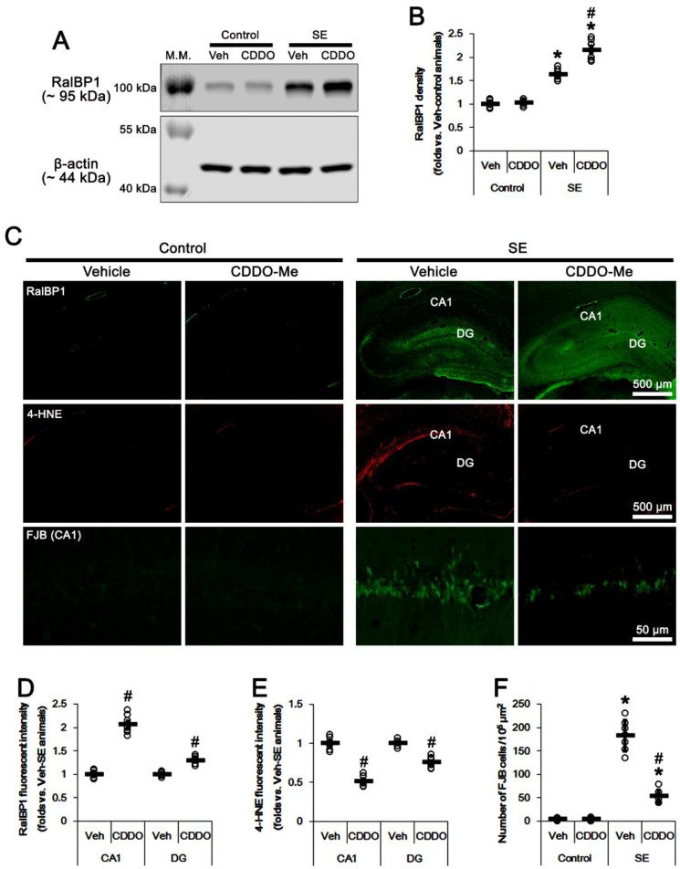
Ras-related protein Ral-A (RalA)-binding protein 1 (RalBP1, also known as Ral-interacting protein of 76 kDa (RLIP76) or Ral-interacting protein 1 (RLIP1 or RIP1)) is involved in the efflux of 4-hydroxynonenal (4-HNE, an end product of lipid peroxidation), as well as mitochondrial fission. In the present study, we found that 2-cyano-3,12-dioxo-oleana-1,9(11)-dien-28-oic acid methyl ester (CDDO-Me) attenuated CA1 neuronal death and aberrant mitochondrial elongations in these neurons coupled with enhanced RalBP1 expression and reduced 4-HNE levels following status epilepticus (SE). RalBP1 knockdown did not affect mitochondrial dynamics and CA1 neuronal death under physiological and post-SE conditions. Following SE, however, cotreatment of RalBP1 siRNA diminished the effect of CDDO-Me on 4-HNE levels, mitochondrial hyperfusion in CA1 neurons, and CA1 neuronal death. These findings indicate that CDDO-Me may ameliorate CA1 neuronal death by facilitating RalBP1-mediated 4-HNE efflux and mitochondrial fission following SE. Therefore, our findings suggest that increased RalBP1 expression/activity may be one of the considerable targets to protect neurons from SE.

Keywords: FJB; mitochondrial dynamics; mitochondrial elongation; mitochondrial fragmentation; oxidative stress; seizure.


Conflict of interest statement

The authors declare no conflict of interest. The funders had no role in the design of the study; in the collection, analyses, or interpretation of data; in the writing of the manuscript, or in the decision to publish the results.

Figures

Figure 1
Figure 1
Effects of CDDO-Me on pilocarpine (PILO)-induced seizure activity. CDDO-Me does not alter seizure susceptibility to pilocarpine. (A) Representative EEG traces and frequency/amplitude maps following pilocarpine treatment. (B) Quantification of the total EEG power (seizure intensity) in response to pilocarpine (mean ± S.E.M.; n = 7; repeated-measures ANOVA).
Figure 2
Figure 2
Effects of CDDO-Me on RalBP1 expression, 4-HNE signals, and CA1 neuronal death following SE. As compared to vehicle, CDDO-Me increases RalBP1 protein levels with reduced 4-HEN signals. CDDO-Me also attenuates SE-induced CA1 neuronal death. (A) Representative Western blots of RalBP1 protein levels in the whole hippocampus (M.W. marker, molecular weight marker). (B) Quantification of RalBP1 protein levels based on Western blot data. (C) Representative photos of RalBP1, 4-HNE, and Fluoro-Jade B (FJB) staining images. SE increases RalBP1 levels in the dentate gyrus (DG), but not in the CA1 region, while it induces 4-HNE accumulation in the CA1 region. CDDO-Me ameliorates SE-induced CA1 neuronal death with increased RalBP1 protein levels and reduced 4-HEN signals. (D,E) Quantification of RalBP1 and 4-HNE fluorescent intensity (mean ± S.E.M.; # p < 0.05 vs. and vehicle; n = 7; Student t-test). (F) Quantification of the number of FJB-positive CA1 neurons following SE (mean ± S.D.; *,# p < 0.05 vs. control animals and vehicle; n = 7; Student t-test). Open circles indicate each individual value. Horizontal bars indicate mean value.
Figure 3
Figure 3
Effects of CDDO-Me on mitochondrial dynamics and RalBP1 expression following SE. As compared to vehicle, CDDO-Me facilitates mitochondrial fission in CA1 neurons under physiological and post-SE conditions. (A) Representative photos of mitochondria (Mito) and RalBP1 expression in CA1 neurons. (B) Quantification of mitochondrial elongation following SE (mean ± S.E.M.; *,# p < 0.05 vs. control animals and vehicle; n = total 25 cells in each group; one-way ANOVA). (C) Quantification of the cumulative area/perimeter ratio and the form factor following SE (mean ± S.E.M.; *,# p < 0.05 vs. control animals and vehicle; n = total 25 cells in each group; one-way ANOVA).
Figure 4
Figure 4
Effects of control siRNA and RalBP1 siRNA on pilocarpine (PILO)-induced seizure susceptibility. RalBP1 knockdown does not influence seizure susceptibility to pilocarpine. (A) Representative EEG traces and frequency/amplitude maps following pilocarpine treatment. (B) Quantification of the total EEG power (seizure intensity) following pilocarpine injection (mean ± S.E.M.; n = 7; repeated-measures ANOVA).
Figure 5
Figure 5
Effects of RalBP1 knockdown on mitochondrial dynamics following SE. RalBP1 siRNA reduces RalBP1 protein levels under physiological and post-SE conditions. However, RalBP1 siRNA does not affect mitochondrial dynamics but aggravates SE-induced CA1 neuronal death. (A) Representative Western blots of RalBP1 protein levels in the whole hippocampus (M.W. marker, molecular weight marker). (B) Quantification of RalBP1 protein levels based on Western blot data. Open circles indicate each individual value. Horizontal bars indicate mean value (mean ± S.E.M.; *,# p < 0.05 vs. control animals and control siRNA; n = 7; one-way ANOVA). (C) Representative photos of mitochondria (Mito) and RalBP1 expression in CA1 neurons. (D) Quantification of mitochondrial elongation index following SE (mean ± S.E.M.). (E) Quantification of the cumulative area/perimeter ratio and the form factor following SE (mean ± S.E.M.). (F) Quantification of the number of FJB-positive CA1 neurons following SE (mean ± S.D.). Open circles indicate each individual value. Horizontal bars indicate mean value (n = 7).
Figure 6
Figure 6
Effects of cotreatment of RalBP1 siRNA with CDDO-Me (CDDO) on pilocarpine (PILO)-induced seizure activity. RalBP1 siRNA does not change the seizure susceptibility to pilocarpine. However, RalBP1 siRNA reduces RalBP1 upregulation induced by CDDO-Me under physiological and post-SE conditions. (A) Representative EEG traces and frequency/amplitude maps following pilocarpine injection. (B) Quantification of the total EEG power (seizure intensity) in response to pilocarpine (mean ± S.E.M.; n = 7; repeated-measures ANOVA). (C) Representative Western blots of RalBP1 protein levels in the whole hippocampus (M.W. marker, molecular weight marker). (D) Quantification of RalBP1 protein levels based on Western blot data. Open circles indicate each individual value. Horizontal bars indicate mean value (mean ± S.E.M.; *,# p < 0.05 vs. control animals and control siRNA; n = 7; one-way ANOVA).
Figure 7
Figure 7
Effects of cotreatment of RalBP1 siRNA on CDDO-Me-induced mitochondrial fission and RalBP1 expression following SE. RalBP1 siRNA cotreatment abrogates the effect of CDDO-Me on mitochondrial fission, 4-HNE accumulation, and neuronal death in CA1 neurons following SE. (A) Representative photos of mitochondria (Mito), RalBP1 expression, 4-HNE accumulation, and SE-induced neuronal damage in CA1 neurons. (B) Quantification of mitochondrial elongation index following SE (mean ± S.E.M.; *,# p < 0.05 vs. control animals and control siRNA; n = total 25 cells in each group; one-way ANOVA). (C) Quantification of the cumulative area/perimeter ratio and the form factor following SE (mean ± S.E.M.; *,# p < 0.05 vs. control animals and control siRNA; n = total 25 cells in each group; one-way ANOVA). (D) Quantification of 4-HNE fluorescent intensity (mean ± S.E.M.; *,# p < 0.05 vs. control animals and control siRNA; n = 7; Student t-test). (E) Quantification of the number of FJB-positive CA1 neurons following SE (mean ± S.D.; *,# p < 0.05 vs. control animals and control siRNA; n = 7; Student t-test). Open circles indicate each individual value. Horizontal bars indicate mean value.

References

    1. Detmer S.A., Chan D.C. Functions and dysfunctions of mitochondrial dynamics. Nat. Rev. Mol. Cell. Biol. 2007;8:870–879. doi: 10.1038/nrm2275. - DOI - PubMed
    1. Kang T.C. Nuclear Factor-Erythroid 2-Related Factor 2 (Nrf2) and Mitochondrial Dynamics/Mitophagy in Neurological Diseases. Antioxidants. 2020;9:617. doi: 10.3390/antiox9070617. - DOI - PMC - PubMed
    1. Kashatus D.F., Lim K.H., Brady D.C., Pershing N.L., Cox A.D., Counter C.M. RALA and RALBP1 regulate mitochondrial fission at mitosis. Nat. Cell Biol. 2011;13:1108–1115. doi: 10.1038/ncb2310. - DOI - PMC - PubMed
    1. Shorvon S., Sen A. What is status epilepticus and what do we know about its epidemiology? Seizure. 2020;75:131–136. doi: 10.1016/j.seizure.2019.10.003. - DOI - PubMed
    1. Bauman K., Devinsky O. Seizure Clusters: Morbidity and Mortality. Front. Neurol. 2021;12:636045. doi: 10.3389/fneur.2021.636045. - DOI - PMC - PubMed