. 2022 May 23;11(5):1030.
doi: 10.3390/antiox11051030.

Identification of Modulators of the C. elegans Aryl Hydrocarbon Receptor and Characterization of Transcriptomic and Metabolic AhR-1 Profiles

Affiliations

Identification of Modulators of the C. elegans Aryl Hydrocarbon Receptor and Characterization of Transcriptomic and Metabolic AhR-1 Profiles

Lucie Larigot et al. Antioxidants (Basel). .

Abstract

The Aryl hydrocarbon Receptor (AhR) is a xenobiotic sensor in vertebrates, regulating the metabolism of its own ligands. However, no ligand has been identified to date for any AhR in invertebrates. In C. elegans, the AhR ortholog, AHR-1, displays physiological functions. Therefore, we compared the transcriptomic and metabolic profiles of worms expressing AHR-1 or not and investigated the putative panel of chemical AHR-1 modulators. The metabolomic profiling indicated a role for AHR-1 in amino acids, carbohydrates, and fatty acids metabolism. The transcriptional profiling in neurons expressing AHR-1, identified 95 down-regulated genes and 76 up-regulated genes associated with neuronal and metabolic functions in the nervous system. A gene reporter system allowed us to identify several AHR-1 modulators including bacterial, dietary, or environmental compounds. These results shed new light on the biological functions of AHR-1 in C. elegans and perspectives on the evolution of the AhR functions across species.

Keywords: Aryl hydrocarbon Receptor; Caenorhabditis elegans; metabolomics; modulators; transcriptomics.


Conflict of interest statement

The authors declare no conflict of interest.

Figures
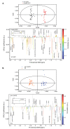
Figure 1
Figure 1
OPLS model discriminating WT-GFP and mutant-GFP (A) or N2 and ahr-1(ia03) (B). Top: Score plots. Bottom: Loading plots. (A) R2 = 0.783, Q2 = 0.973. mutant-GFP is associated with high levels of phenylalanine, cystathionine, lysine, b-alanine, glutamine, tyrosine, valine, leucine and isoleucine, and low levels of allantoin, trehalose, phosphocholine and glycerophosphocholine, glutamate, cyclic fatty acids and glyceryl of lipids, by comparison to WT-GFP. (B) R2 = 0.903, Q2 = 0.93. ahr-1(ia03) is associated with high levels of phenylalanine, glycerol, betaine, cystathionine, lysine, asparagine, tyrosine, valine, leucine and isoleucine, and low levels of allantoin, trehalose, glycerophosphocholine, phosphocholine, cyclic fatty acids and glyceryl of lipids, by comparison to N2.
Figure 2
Figure 2
Requirement of AHR-1 and AHA-1 for transcriptional activity. (A) Structure of AHR-1 WT, ahr-1(ju145) and ahr-1(ia03) proteins. The 602 amino acids AHR-1 WT protein is truncated in ahr-1(ju145) as a result of a C to T point mutation that leads to a stop codon (Huang et al., 2004). The ahr-1(ia03) mutant allele contains a 1517 bp deletion from 205 bp 5′ of exon 4 to 30 bp 5′ of exon 8 that results in a frameshift and a premature stop codon (Qin and Powell-Coffman, 2004). (B) The requirement of the two partners is examined in the screening model. The Cos-7 cells transfection is carried out in different conditions with 5 ng/well of ahr-1 (WT or mutants) and aha-1 plasmids and without FBS: either with the empty vector (pcDNA3) or with only one of the two partners (AHA-1, ahr-1(ju145) or AHR-1 WT) and with the two partners (ahr-1(ju145):AHA-1 and AHR-1 WT:AHA-1). Six independent experiments were performed in duplicate, error bars represent SD. Friedman test with Dunn’s multiple comparisons post-test * p-value < 0.05, *** p-value < 0.001.
Figure 3
Figure 3
Comparison between Cos-7 cells transfected with C. elegans ahr-1 or human ahr. Cos-7 cells were transfected with the empty vector (pcDNA3) or with C. elegans ahr-1/aha-1 or with human ahr/arnt. After transfection and culture for 24 h in 0% FBS medium, cells were treated for 24 h with 10 nM of TCDD. The relative activity represents the Firefly measurement normalized to the Renilla measurement for each condition. Three independent experiments were performed in duplicate, error bars represent SD. ANOVA followed with comparison test of each column with the control column (pcDNA3—No treatment) *** p-value < 0.001.
Figure 4
Figure 4
AHR-1 basal activity in Cos-7 cells. Different plasmid concentrations and FBS conditions were tested during the screening model optimization. (A) Comparison between the transfection of the empty vector (pcDNA3) and the two partners (ahr-1 and aha-1 plasmids) in Cos-7 cells cultured with 10% FBS. (B) Comparison between the transfection of ahr-1 and aha-1 plasmids in Cos-7 cells cultured either in 0% FBS or in 10% FBS. The relative activity represents the measurement of the Firefly activity normalized to the Renilla measurement for each condition. Six independent experiments were performed in duplicate, error bars represent SD. ANOVA with Bonferroni’s multiple comparison post-test * p-value < 0.05, *** p-value < 0.001.
Figure 5
Figure 5
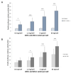
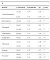
Summary of significant AHR-1 modulators in Cos-7 cells. Positive (A) and negative (B) modulator’s concentrations are represented with the dependent-AHR-1 fold induction of Firefly luciferase. Example of a positive (C), and negative (D) modulator dependent concentration effect on AHR-1 activity. Six independent experiments were performed in duplicate, fold induction is standardized to the vehicle which is 1. Statistical significances relative to the vehicle were examined: * p-value < 0.05; ** p-value < 0.01; *** p-value < 0.001. (E) Molecular structure of the positive (green) and negative (red) modulators of AHR-1.
Figure 5
Figure 5
Summary of significant AHR-1 modulators in Cos-7 cells. Positive (A) and negative (B) modulator’s concentrations are represented with the dependent-AHR-1 fold induction of Firefly luciferase. Example of a positive (C), and negative (D) modulator dependent concentration effect on AHR-1 activity. Six independent experiments were performed in duplicate, fold induction is standardized to the vehicle which is 1. Statistical significances relative to the vehicle were examined: * p-value < 0.05; ** p-value < 0.01; *** p-value < 0.001. (E) Molecular structure of the positive (green) and negative (red) modulators of AHR-1.
Figure 6
Figure 6
Time course of 3MC, a positive modulator of AHR-1 in Cos-7 cells. After transfection and culture for 24 h in 0% FBS medium, cells were treated for 24 h with 3-methylcholanthrene 5 µM. After 3 h, 5 h, 16 h and 26 h of treatment, cells were lysed, and Firefly luciferase luminescence was read. Six independent experiments were performed in duplicate, error bars represent SD, fold induction is standardized to the vehicle which is 1. Statistical significances relative to the vehicle were examined: ANOVA with Bonferroni’s multiple comparison post-test, * p-value < 0.05.

References

    1. Poland A., Clover E., Kende A.S., DeCamp M., Giandomenico C.M. 3,4,3′,4′-Tetrachloro Azoxybenzene and Azobenzene: Potent Inducers of Aryl Hydrocarbon Hydroxylase|Science. Science. 1976;194:627–630. doi: 10.1126/science.136041. - DOI - PubMed
    1. Poland A., Palen D., Glover E. Tumour Promotion by TCDD in Skin of HRS/J Hairless Mice. Nature. 1982;300:271–273. doi: 10.1038/300271a0. - DOI - PubMed
    1. Whitlock J.P. Genetic and Molecular Aspects of 2,3,7,8-Tetra-Chlorodibenzo-P-Dioxin Action. Annu. Rev. Pharmacol. Toxicol. 1990;30:251–277. doi: 10.1146/annurev.pa.30.040190.001343. - DOI - PubMed
    1. Nguyen L.P., Bradfield C.A. The Search for Endogenous Activators of the Aryl Hydrocarbon Receptor. Chem. Res. Toxicol. 2008;21:102–116. doi: 10.1021/tx7001965. - DOI - PMC - PubMed
    1. Hoffman E., Reyes H., Chu F., Sander F., Conley L., Brooks B., Hankinson O. Cloning of a Factor Required for Activity of the Ah (Dioxin) Receptor. Science. 1991;252:954–958. doi: 10.1126/science.1852076. - DOI - PubMed