Case Reports
. 2022 Apr 29;12(5):574.
doi: 10.3390/brainsci12050574.

Duplication of the Pituitary Gland: CT, MRI and DTI Findings and Updated Review of the Literature

Affiliations
Case Reports

Duplication of the Pituitary Gland: CT, MRI and DTI Findings and Updated Review of the Literature

Bettina L Serrallach et al. Brain Sci. .

Abstract

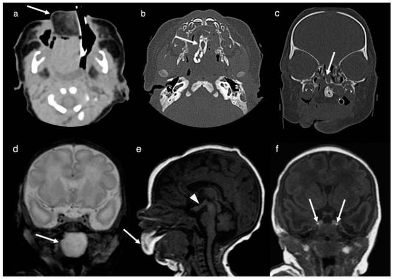
Duplication of the pituitary gland (DPG) is an extremely rare malformation. DPG is associated with a wide variety of midline and central nervous system malformations (DPG-plus syndrome). We present the computed tomography (CT), magnetic resonance imaging (MRI) and diffusion tensor imaging (DTI) findings of a rare case of DPG with associated tuberomammillary fusion resulting in a hypothalamic mass-like configuration, oropharyngeal teratoma, cleft palate, hypertelorism, duplicated/broad sella, duplication/low bifurcation of the basilar artery, and craniovertebral midline anomalies. Qualitative interpretation of DTI yielded normal white matter organization of the brain. The duplication of the prechordal plate and the rostral end of the notochordal plate/notochord is thought to be the main factor leading to a duplication of the pituitary primordium and resulting in the formation of two morphologically normal glands. The time of induction of the teratogenic influence, the extent of the prechordal plate and notochordal plate/notochord abnormalities, and the faulty interactions are believed to be the reason for the wide spectrum of associated midline abnormalities.

Keywords: DPG-plus syndrome; computed tomography (CT); diffusion tensor imaging (DTI); duplication of the pituitary gland (DPG); embryology; magnetic resonance imaging (MRI).


Conflict of interest statement

The authors declare no conflict of interest.

Figures

Figure 1
Figure 1
Images obtained at the second day of life. Axial CT-image (a), axial CT-image (bone algorithm, (b)), coronal CT-image (bone algorithm, (c)), coronal T2-weighted MR-image (d) and sagittal T1-weighted MR-image (e) show a heterogeneous mass centered in the oral cavity and protruding anteriorly through the mouth. The mature teratoma is composed out of fatty tissue anteriorly (arrows in (a,e)), a mineralized/bony component in the midline (arrow in (b)) and a cystic component posteriorly within the naso-/oropharynx (arrow in (d)). A cleft palate with corresponding defect in the hard palate can be seen in (c) (arrow). The tuberomammillary fusion is shown in (e) (arrowhead). Coronal T1-weighted MR-image (f) documents a complete duplication of the pituitary gland including the pituitary stalk. The bright spots of the duplicated posterior pituitary are clearly depicted (arrows in (f)).
Figure 2
Figure 2
Brain MRI images obtained at 8 years of age. Coronal T2-weighted short-tau inversion recovery (STIR; (a)), axial T2-weighted (b), coronal fat saturated T1+C-weighted (c) and axial T1+C-weighted (d) MR-images demonstrate the complete duplication of the pituitary gland including the pituitary stalk (arrows in (ad)). Sagittal T1-weighted MR-image (e) documents the tuberomammillary fusion (arrow). Coronal T2-weighted STIR image (f) shows the duplication/low bifurcation of the basilar artery (arrow) at the midpons level with the distal basilar artery supplying the superior cerebellar and posterior cerebral arteries.
Figure 3
Figure 3
CT obtained at the age of 8 years. Coronal CT-image (bone algorithm; (a)), axial CT-3D reconstruction (b), axial CT-image (bone window, (c)) and axial CT-3D reconstruction (d). Images demonstrate a broadened/duplicated odontoid process (arrowhead in (a)), a bony fusion of a midline ossicle with the clivus (arrow in (a) and arrowhead in (b)), a broadened base of C2 (arrow in (c)), a congenital absence of the anterior arch of the atlas (c), an incomplete fusion of the posterior synchondrosis (arrowhead in (c)) and a broad and shallow sella turcica, which is divided into two fossae by a midline osseous ridge (arrowhead in (b) and close up in (d)).
Figure 4
Figure 4
DTI obtained at 8 years of age. Axial (a), and sagittal (b) color-coded fractional anisotropy (FA) map showing qualitative normal white matter organization of the brain. Coronal fiber tractography superimposed on color-coded FA map (c) displays the normal course of the corticospinal tracts (CST) on both sides. Axial color-coded FA map at the level of the middle pons (close up, (d)). The principal diffusion direction within the areas of high degree anisotropic diffusion is color-coded in red (left to right diffusion), blue (superior-inferior diffusion) and green (anterior-posterior diffusion). At the level of the middle pons (d), the CST (anteriorly) and the medial lemniscus (ML) (posteriorly) can be appreciated as blue vertical structures. Between the CST and ML, the transverse pontine fibers (TPF) can be seen as a red structure. The medial cerebellar peduncle (MCP) can be detected as a green structure laterally to the brainstem. The superior cerebellar peduncle (SCP) is shown as a light blue-green structure (due to a slightly vertical orientation) medially to the MCP.

References

    1. Accornero S., Danesino C., Bastianello S., D’Errico I., Guala A., Chiovato L. Duplication of the pituitary stalk in a patient with a heterozygous deletion of chromosome 14 harboring the thyroid transcription factor-1 gene. J. Clin. Endocrinol. Metab. 2010;95:3595–3596. doi: 10.1210/jc.2010-0621. - DOI - PubMed
    1. Ahmed A., Ehsan A.N., Mubarak F., Shamim M.S., Batool M., Banu S. Unique Presentation of Duplication of the Pituitary Gland-Plus Syndrome. AACE Clin. Case Rep. 2020;6:e357–e360. doi: 10.4158/ACCR-2019-0487. - DOI - PMC - PubMed
    1. Akin L., Kendirci M., Doganay S., Kurtoglu S., Tucer B., Coskun A. Pituitary duplication: A rare cause of precocious puberty. Childs Nerv. Syst. 2011;27:1157–1160. doi: 10.1007/s00381-011-1443-8. - DOI - PubMed
    1. Azurara L., Marcal M., Vieira F., Tuna M.L. DPG-plus syndrome: New report of a rare entity. BMJ Case Rep. 2015;2015:212416. doi: 10.1136/bcr-2015-212416. - DOI - PMC - PubMed
    1. Burke M., Zinkovsky S., Abrantes M.A., Riley W. Duplication of the hypophysis. Pediatr. Neurosurg. 2000;33:95–99. doi: 10.1159/000028983. - DOI - PubMed

Publication types

LinkOut - more resources