. 2022 May 11;12(10):1236.
doi: 10.3390/ani12101236.

Occurrence of Fibropapillomatosis in Green Turtles (Chelonia mydas) in Relation to Environmental Changes in Coastal Ecosystems in Texas and Florida: A Retrospective Study

Affiliations

Occurrence of Fibropapillomatosis in Green Turtles (Chelonia mydas) in Relation to Environmental Changes in Coastal Ecosystems in Texas and Florida: A Retrospective Study

Costanza Manes et al. Animals (Basel). .

Abstract

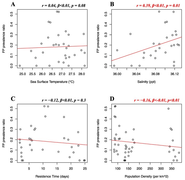
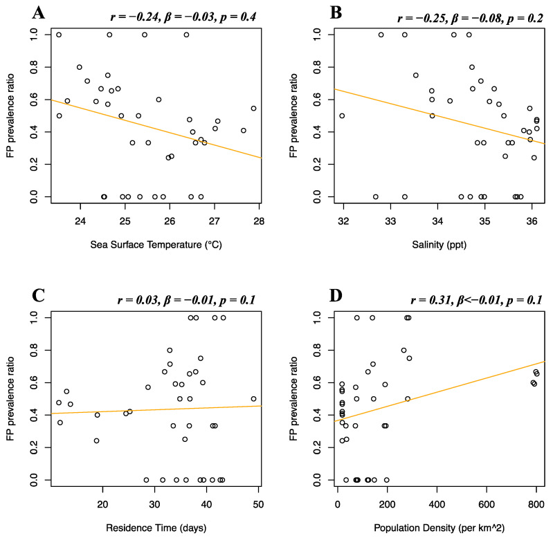
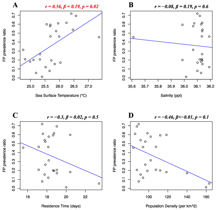
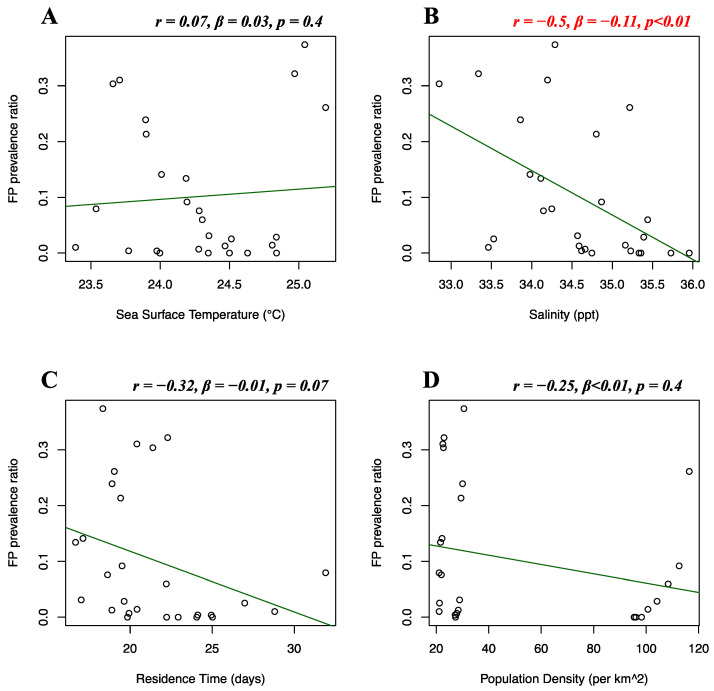
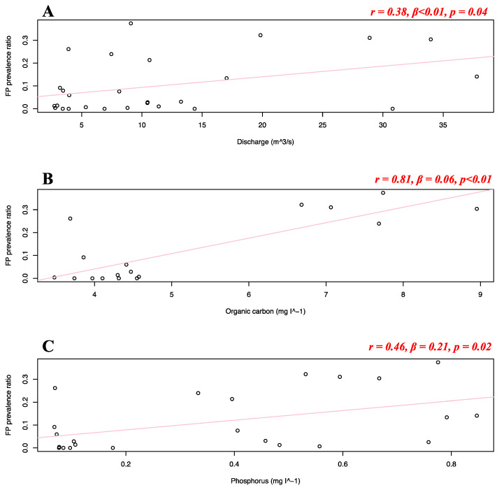
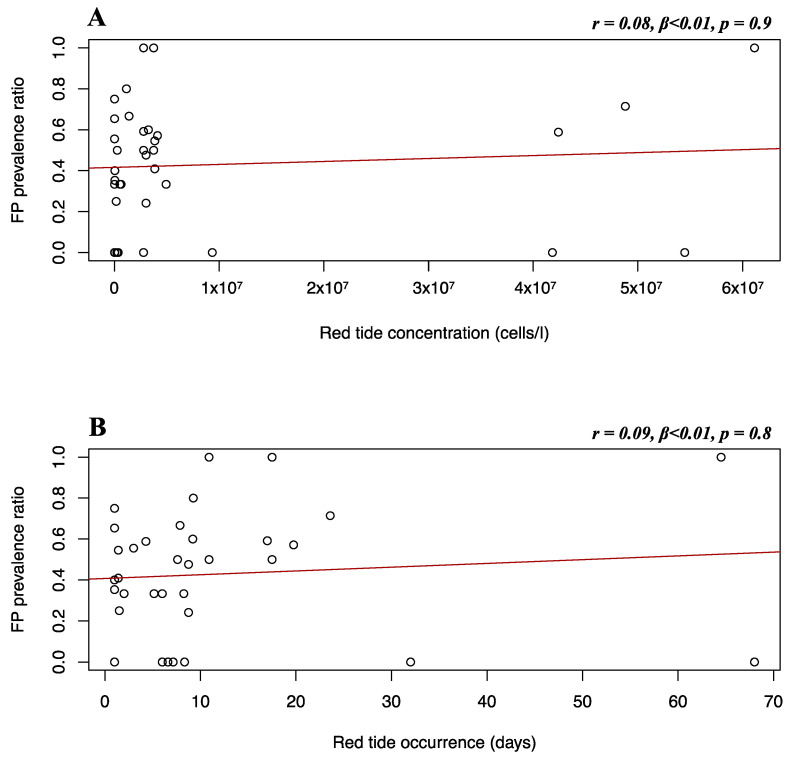
Fibropapillomatosis is a neoplastic disease of marine turtles, with green turtles (Chelonia mydas) being the most affected species. Fibropapillomatosis causes debilitating tumor growths on soft tissues and internal organs, often with lethal consequences. Disease incidence has been increasing in the last few decades and the reason is still uncertain. The potential viral infectious agent of Fibropapillomatosis, chelonid herpesvirus 5, has been co-evolving with its sea turtle host for millions of years and no major mutation linked with increased disease occurrence has been detected. Hence, frequent outbreaks in recent decades are likely attributable to external drivers such as large-scale anthropogenic changes in the green turtle coastal marine ecosystem. This study found that variations in sea surface temperature, salinity, and nutrient effluent discharge from nearby rivers were correlated with an increased incidence of the disease, substantiating that these may be among the significant environmental drivers impacting Fibropapillomatosis prevalence. This study offers data and insight on the need to establish a baseline of environmental factors which may drive Fibropapillomatosis and its clinical exacerbation. We highlight the multifactorial nature of this disease and support the inclusion of interdisciplinary work in future Fibropapillomatosis research efforts.

Keywords: coastal waters; ecosystem change; environmental impact; marine ecology; turtles; viruses; wildlife diseases.


Conflict of interest statement

The authors declare no conflict of interest.

Figures

Figure 1
Figure 1
Timeframe of retrospective study. The orange blocks represent the timeframes of FP prevalence data shared by 3rd party authors or available in published papers for the different locations (1980–1998 for Florida stranded FP dataset [20]; 1983–2006 for Florida in-water FP dataset [50,51]; 2010–2018 for Texas FP dataset [14]). Matching timeframe of availability of predictor variables is reported for each group. Absence of predictor variables block in a given location (i.e., red tide data in Florida in-water locations) corresponds to NAs. Not all FP data could be utilized for the purposes of our study. Statistical analyses were run solely on the space and time matching timeframes from the earliest available timeframe after demographic data availability (i.e., patterns of Florida in-water FP prevalence versus environmental and demographic predictors from 1995 to 2006, see Section 2.2.1).
Figure 2
Figure 2
(A) Distribution of the surveyed turtles between 1980 and 1998 [20]; (B) distribution and prevalence of surveyed turtles affected by FP in Texas from 2010 to 2018 (adapted from Shaver et al., 2019) [14].
Figure 3
Figure 3
Distribution of the watersheds we identified in the study areas to divide the riverine water quality data and the population densities (orange polygons), and the areas we considered for the extraction of the environmental data from the HYCOM hydrostatic circulation model (blue polygons) in Florida (A) and Texas (B). Distribution of the USGS stations (green dots) in Florida (C) and Texas (D), where data was available in the considered timeframe (see Figure 1).
Figure 4
Figure 4
Heat maps representing the number of observations (A), and the average concentration in cells/liter (B) of K. brevis collected by FWC and FWRI (Fish and Wildlife Conservation Commission—Fish and Wildlife Research Institute) in the time frame where the environmental data were available (from 1987 through 1998, see Figure 1). The heat maps are represented using a regular grid. The dimension of the cells constituting the grid is 10 km × 10 km.
Figure 5
Figure 5
Florida Atlantic coast stranded dataset FP prevalence and environmental and demographic variables. Scatterplots with fitted regression lines representing the logistic regression output from the response variables versus predictor variables in the Florida Atlantic coast stranded dataset. The subplots show FP prevalence ratio on the y-axis versus sea surface temperature (A), salinity (B), residence time (C), and human population density (D) on the x-axis. Pearson correlation (r), beta-coefficient (β), and p-value (p) are reported for each predictor variable on the top right of the respective plot. Significant relationships are highlighted in red.
Figure 6
Figure 6
Florida Gulf coast stranded dataset FP prevalence and environmental and demographic variables. Scatterplots with fitted regression lines representing the logistic regression output from the response variables versus predictor variables in the Florida Gulf coast stranded dataset. The subplots show FP prevalence ratio on the y-axis versus sea surface temperature (A), salinity (B), residence time (C), and human population density (D) on the x-axis. Pearson correlation (r), beta-coefficient (β), and p-value (p) are reported for each predictor variable on the top right of the respective plot.
Figure 7
Figure 7
Florida in-water dataset FP prevalence and environmental and demographic variables. Scatterplots with fitted regression lines representing the logistic regression output from the response variables versus predictor variables in the Florida in-water dataset. The subplots show FP prevalence ratio on the y-axis versus sea surface temperature (A), salinity (B), residence time (C), and human population density (D) on the x-axis. Pearson correlation (r), beta-coefficient (β), and p-value (p) are reported for each predictor variable on the top right of the respective plot. Significant relationships are highlighted in red.
Figure 8
Figure 8
Texas FP prevalence and environmental and demographic variables. Scatterplots with fitted regression lines representing the logistic regression output from the response variables versus predictor variables in the Texas dataset. Subplots show FP prevalence ratio on the y-axis versus sea surface temperature (A), salinity (B), residence time (C), and population density (D) on the x-axis. Pearson correlation (r), beta-coefficient (β), and p-value (p) are reported for each predictor variable on the top right of the respective plot. Significant relationships are highlighted in red.
Figure 9
Figure 9
Texas dataset FP prevalence and discharge and nutrients variables. Scatterplots with fitted regression lines representing the logistic regression output from the response variables versus predictor variables in the Texas dataset: FP prevalence ratio on the y-axis versus discharge (A), organic carbon (B), and phosphorus (C). Pearson correlation (r), beta-coefficient (β), and p-value (p) are reported for each predictor variable on the top right of the respective plot. Significant relationships are highlighted in red.
Figure 10
Figure 10
Florida Gulf coast stranded dataset FP prevalence and red tide variables. Scatterplots with fitted regression lines representing the logistic regression output from the response variables versus predictor variables in the Florida Gulf coast stranded dataset: FP prevalence ratio on the y-axis versus red tide concentration (A) and red tide occurrence (B) on the x-axis. Pearson correlation (r), beta-coefficient (β), and p-value (p) are reported for each predictor variable on the top right of the respective plot.

References

    1. Jones K., Ariel E., Burgess G., Read M. A review of fibropapillomatosis in Green turtles (Chelonia mydas) Vet. J. 2016;212:48–57. doi: 10.1016/j.tvjl.2015.10.041. - DOI - PubMed
    1. Duffy D.J., Schnitzler C., Karpinski L., Thomas R., Whilde J., Eastman C., Yang C., Krstic A., Rollinson D., Zirkelbach B., et al. Sea turtle fibropapilloma tumors share genomic drivers and therapeutic vulnerabilities with human cancers. Commun. Biol. 2018;1:1–13. doi: 10.1038/s42003-018-0059-x. - DOI - PMC - PubMed
    1. Smith G.M., Coates C.W. Fibro-epithelial growths of the skin in large marine turtles, Chelonia mydas (Linnaeus) Zoologica. 1938;23:93–96. doi: 10.5962/p.203654. - DOI
    1. Hirama S., Ehrhart L.M. Description, prevalence and severity of green turtle fibropapillomatosis in three developmental habitats on the east coast of Florida. Florida Sci. 2007;70:435–448.
    1. Lawrance M.F., Mans K.L., Sutton E., Savage A.E. Molecular evolution of fi bropapilloma-associated herpesviruses infecting juvenile green and loggerhead sea turtles. Virology. 2018;521:190–197. doi: 10.1016/j.virol.2018.06.012. - DOI - PubMed

LinkOut - more resources