Review
. 2022 Apr 19;12(5):600.
doi: 10.3390/biom12050600.

Advances towards Understanding the Mechanism of Action of the Hsp90 Complex

Affiliations
Review

Advances towards Understanding the Mechanism of Action of the Hsp90 Complex

Chrisostomos Prodromou et al. Biomolecules. .

Abstract

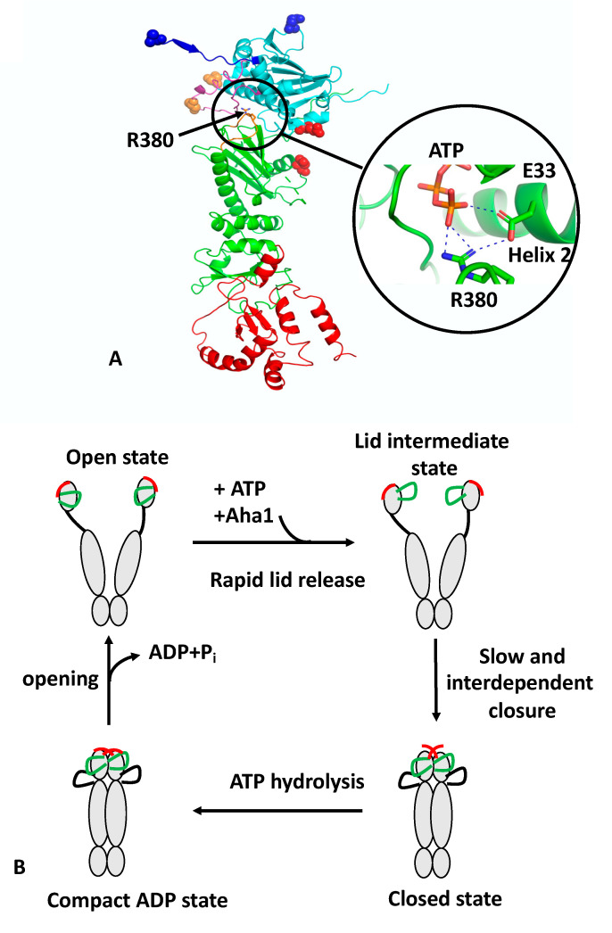
Hsp90 (Heat Shock Protein 90) is an ATP (Adenosine triphosphate) molecular chaperone responsible for the activation and maturation of client proteins. The mechanism by which Hsp90 achieves such activation, involving structurally diverse client proteins, has remained enigmatic. However, recent advances using structural techniques, together with advances in biochemical studies, have not only defined the chaperone cycle but have shed light on its mechanism of action. Hsp90 hydrolysis of ATP by each protomer may not be simultaneous and may be dependent on the specific client protein and co-chaperone complex involved. Surprisingly, Hsp90 appears to remodel client proteins, acting as a means by which the structure of the client protein is modified to allow its subsequent refolding to an active state, in the case of kinases, or by making the client protein competent for hormone binding, as in the case of the GR (glucocorticoid receptor). This review looks at selected examples of client proteins, such as CDK4 (cyclin-dependent kinase 4) and GR, which are activated according to the so-called 'remodelling hypothesis' for their activation. A detailed description of these activation mechanisms is paramount to understanding how Hsp90-associated diseases develop.

Keywords: ATPase; Aha1; Cdc37; Hsp90; chaperone; co-chaperone; heat shock proteins; immunophilins; kinase; mechanism; p23; steroid hormone receptor; structure.


Conflict of interest statement

The authors declare no conflict of interest.

Figures

Figure 1
Figure 1
PyMol cartoon showing the structure and conformational switches of Hsp90 and its catalytic cycle. (A) Monomeric Hsp90 in the active, N-terminally dimerised closed conformation. Hsp90 consists of three domains: cyan, NTD; green, MD; and red, CTD. Four structural features are shown: blue, the structure involved in the N-terminal β-strand cross-subunit swapping; magenta, the lid-segment involved in lid closure; cyan and green, the NTD and MD domains involved in intra-subunit association; and magenta, the catalytic loop containing Arg 380 (shown as sticks). Attachment points for the fluorophore or Trp are shown as residues in sphere representation: blue, represents the n-terminal β-strand cross-subunit swapping switch pair (normally as a pair between both protomers); red, the NTD and MD intra-subunit association switch; and gold, the lid-closure switch. The close up shows the interaction of Arg 380 with the γ-phosphate of ATP and Glu 33. Dotted blue lines represent salt bridges. (B) The catalytic cycle of Hsp90. The apo state of Hsp90 occupies a heterologous ensemble of open-conformers. The lid (green) and n-terminal β-strand (red) are highly mobile structural elements showing a sub-millisecond reconfiguration time. The binding of ATP to the NTD of Hsp90 leads to a rapid release of the lid segment to an intermediate conformational state, between the so-called open- and closed-lid states. The association of Aha1 can remodel the lid but also preassociates the n- and m-domains for accelerated closure. Cooperative action of conformational switches leads to full closure of the molecular clamp. This involves closure of the lid over the ATP-binding pocket, a cross-subunit swap of the β-strands, and association of the n- and m-domains, which collectively are slow and interdependent. The β-strand swapping is weakly coupled with the other motions. The cycle completes following the hydrolysis of ATP, which leads to a compact, ADP-bound conformation. The hydrolysis of ATP appears to be non-cooperative between the two protomers of Hsp90. Following ATP hydrolysis, Hsp90 then relaxes to an open state, with concomitant release of ADP and inorganic phosphate.
Figure 2
Figure 2
PyMol cartoons showing the interactions of Hsp90, Cdc37 and kinases. (A) The interaction of the MD and CTD of Cdc37 (cyan) with the NTD of Hsp90 (green). The Hsp90 lid is shown in magenta. Arg 167 of Cdc37 forms a salt bridge with Glu 33 of Hsp90. Residues are shown in stick format. (B) The negative stain EM structure of the Hsp90–Cdc37–Cdk4 complex, where Cdk6 was superimposed into the electron density of Cdk4 [31]. (C) The cryo-EM structure of the Hsp90–Cdc37–Cdk4 complex [13]. Note that the MD of Cdc37 is centered above Trp 312. (D) Interaction of the HPNI motif (sticks) of Cdc37 (magenta) with the C-terminal domain of Cdk4 (yellow). (E) Interaction of the NTD of Cdc37 (gold to yellow) with Hsp90 (green and cyan). Interactions are seen to occur with the catalytic loop (magenta) and the long helix of the MD and NTD of Hsp90. Phospho-Ser 13 (pS13) helps to stabilise the N-terminal loop of Cdc37 and forms important interactions with the base of the N-terminal end of the helix–turn–helix of the NTD of Cdc37. Some potential interactions are shown using dashed lines (blue). Blue, the CTD of Cdk4.
Figure 3
Figure 3
PyMol cartoons showing the Hsp90–Aha1 interactions. (A) apo-Hsp90 (yellow and cyan) in complex with two molecules of Aha1 (gold, N-Aha, and salmon, C-Aha1). Blue, amphipathic helix (B) AMPPNP-Hsp90 (yellow and cyan) in complex with two molecules of Aha1 (magenta and salmon), although only one N-terminal domain of Aha1 was observed in the complex. The rotation of the NTD of Aha1 from its apo-Hsp90 (green) to the AMPPNP-Hsp90 location (magenta) is shown. (C) Interaction of segments of Aha1 (magenta, residues 1–10 and gold, residues 154–170) with the NTDs of Hsp90 (yellow and cyan). Grey, surface lid segment of Hsp90. (D) Rotation of the CTD of Aha1 from its apo-Hsp90 (green and cyan) to the AMPPNP-Hsp90 location (salmon and yellow) results in changes of interaction from Phe 328 to Trp 296 of Aha1. (E) Chaperone cycles in response to Aha1 binding.
Figure 4
Figure 4
PyMol cartoons showing the Hsp90-p23 interactions. (A) Interaction of p23 (yellow) with the NTDs of both Hsp90 protomers (green and cyan) and the MDs of one Hsp90 protomer (magenta). (B) Interaction of the C-terminal unstructured segment of Sba1 (green) and p23 (yellow) with the long helix of the MD of Hsp90 (cyan). Numbers in brackets are for the human protein. (C) Comparison of the position of Phe 121 and W124 of Sba1 (wheat), which engage with the middle domain of Hsp90 (not shown) and the gradual engagement of specific Aha1 residues (yellow [apo-Hsp90-Aha1] to orange [Yeast Hsp90-Aha1] to cyan [Hsp90–AMPPNP–Aha1; fully tilted structure]) at similar positions with those from Sba1. Residues between Sba1 and Aha1 more or less overlap as the fully closed complex of the Hsp90–Aha1 complex is formed. Overlap of positions occur for Trp 11 and Leu 66 of Aha1, which mimic the position of Phe 121 and Trp124 of Sba1, respectively. (D) Interaction of the C-terminal helix 7 of FKBP51 (salmon) with the C-terminal hydrophobic pocket at the C-terminal dimer interface of Hsp90 (green and cyan). Interacting residues are shown in stick format and polar interactions as dotted blue lines.
Figure 5
Figure 5
PyMol cartoons showing interactions within the Hsp90–Hsp70–HOP–GR loading complex. (A) Interface 1 and 2 of the Hsp90–Hsp70 interaction. A β-strand (β) from the outer face of the MD β-sheet of Hsp90 (green) inserts itself into the cleft between the subdomains IA and IIA of Hsp70 (cyan IIA and yellow, IA) in interface 1. This cleft is only available in the ADP state of Hsp70. Interface 2 is shown on the right-hand side of the panel. (B) The interaction of HOP and Hsp70 within the Hsp90–Hsp70–Hop-GR complex. Only three domains of Hop were visible, TPR2A, TPR2B and the DP2 domain, which appear to be fully sufficient for full GR activation. The binding of HOP is essential to maintaining the semi-closed conformation of Hsp90 and also for assembling the two bound Hsp70 molecules. Salmon, HOP; cyan and green, Hsp90 dimer; red, lid-segment of Hsp90; blue, N-terminal segments of Hsp90; and slate, Hsp70. (C) Interaction of the DP2 domain of HOP with the conserved client-protein binding-site residues of Hsp90. Green, Hsp90 dimer and salmon, HOP. (D) GR domain interactions with the Hsp90 complex. Yellow, GR; cyan and green, Hsp90 dimer; and slate, Hsp70. (E) Interaction of helix 1 and the pre-helix-1 segment of GR with DP2 and Hsp70, respectively. Yellow, GR; cyan, Hsp90; green, Hsp70 substrate-binding domain; and salmon, HOP DP2 domain.
Figure 6
Figure 6
PyMol cartoon of the interactions of the Hsp90-p23-GR complex. (A) The pre-helix-1 held within the hydrophobic lumen of Hsp90. Yellow, GR; and cyan and green, Hsp90 dimer. (B) GR LBD (yellow) interacts with residues Trp 320 and Phe 349 from the adjacent Hsp90 protomer (green). (C) Helix 1 of GR packing against the amphipathic helix of Hsp90. Yellow, GR; cyan, Hsp90; and substrate binding sites of Hsp90 are shown in gold and slate (amphipathic helix). (D) The C-terminal tail of p23 (magenta) interacts with the long helix of the MD of Hsp90 (cyan and green) and the catalytic Arg 400 is in contact with the bound nucleotide (stick representation). (E) A relay of polar interactions between the C-terminal tail of p23 (magenta) and Hsp90 (cyan and green) and GR (yellow). (F,G) The extreme C-terminus helix of p23 (magenta) interacts with GR (yellow).
Figure 7
Figure 7
The proposed GR activation cycle. 1 to 4 represents the steps of the cycle. Grey, GR LBD; magenta and yellow, Hsp70; green and cyan, Hsp90 dimer; salmon, HOP; and orange, p23.

References

    1. Biebl M.M., Buchner J. Structure, Function, and Regulation of the Hsp90 Machinery. Cold Spring Harb. Perspect. Biol. 2019;11:a034017. doi: 10.1101/cshperspect.a034017. - DOI - PMC - PubMed
    1. Prodromou C. Mechanisms of Hsp90 regulation. Biochem. J. 2016;473:2439–2452. doi: 10.1042/BCJ20160005. - DOI - PMC - PubMed
    1. Prodromou C. The “active life” of Hsp90 complexes. Biochim. Biophys. Acta. 2012;1823:614–623. doi: 10.1016/j.bbamcr.2011.07.020. - DOI - PMC - PubMed
    1. Prodromou C., Morgan R.M.L. Tuning the ATPase Activity of Hsp90. In: Chakraborti S., Dhalla N.S., editors. Regulation of Ca2+-ATPases, V-ATPases and F-ATPases. Advances in Biochemistry in Health and Disease. Volume 14. Springer; Cham, Switzerland: 2016. pp. 469–490.
    1. Meyer P., Prodromou C., Liao C., Hu B., Roe S.M., Vaughan C.K., Vlasic I., Panaretou B., Piper P.W., Pearl L.H. Structural basis for recruitment of the ATPase activator Aha1 to the Hsp90 chaperone machinery. EMBO J. 2004;23:1402–1410. doi: 10.1038/sj.emboj.7600141. - DOI - PMC - PubMed

MeSH terms

LinkOut - more resources