

Star Polymers as Non-Viral Carriers for Apoptosis Induction
- PMID: 35625536
- PMCID: PMC9139127
- DOI: 10.3390/biom12050608
Star Polymers as Non-Viral Carriers for Apoptosis Induction
Abstract
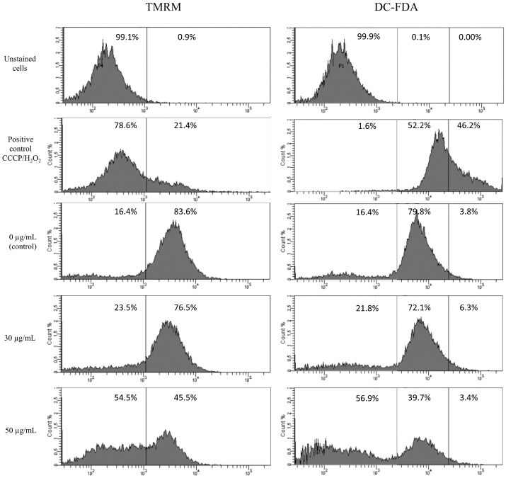
Apoptosis is a widely controlled, programmed cell death, defects in which are the source of various diseases such as neurodegenerative diseases as well as cancer. The use of apoptosis in the therapy of various human diseases is of increasing interest, and the analysis of the factors involved in its regulation is valuable in designing specific carriers capable of targeting cell death. Highly efficient and precisely controlled delivery of genetic material by low-toxic carriers is one of the most important challenges of apoptosis-based gene therapy. In this work, we investigate the effect of the star polymer with 28 poly(N,N'-dimethylaminoethyl methacrylate) arms (STAR) on human cells, according to its concentration and structure. We show that star polymer cytotoxicity increases within its concentration and time of cells treatment. Except for cytotoxic effect, we observe morphological changes such as a shrinkage, loss of shape and begin to detach. We also prove DNA condensation after star polymer treatment, one of the most characteristic feature of apoptosis. The results indicate that the use of STAR triggers apoptosis in cancer cells compared to various normal cells, what makes these nanoparticles a promising drug in therapeutic strategy, which targets apoptosis. We demonstrate highlighting potential of star polymers as an innovative tool for anti-cancer therapy.
Keywords: apoptosis; cross-membrane transport; delivery systems; star polymers vectors.
Conflict of interest statement
The authors declare no conflict of interest.
Figures


Similar articles
-
Star-shaped poly(2-aminoethyl methacrylate)s as non-viral gene carriers: Exploring structure-function relationship.Colloids Surf B Biointerfaces. 2019 Sep 1;181:721-727. doi: 10.1016/j.colsurfb.2019.06.029. Epub 2019 Jun 15. Colloids Surf B Biointerfaces. 2019. PMID: 31228855
-
Nonviral Plasmid DNA Carriers Based on N,N'-Dimethylaminoethyl Methacrylate and Di(ethylene glycol) Methyl Ether Methacrylate Star Copolymers.Biomacromolecules. 2015 Oct 12;16(10):3275-85. doi: 10.1021/acs.biomac.5b00948. Epub 2015 Sep 24. Biomacromolecules. 2015. PMID: 26375579
-
A Rationally Optimized Nanoparticle System for the Delivery of RNA Interference Therapeutics into Pancreatic Tumors in Vivo.Biomacromolecules. 2016 Jul 11;17(7):2337-51. doi: 10.1021/acs.biomac.6b00185. Epub 2016 Jun 28. Biomacromolecules. 2016. PMID: 27305597
-
Drug carriers with star polymer structures.Physiol Res. 2018 Oct 30;67(Suppl 2):S293-S303. doi: 10.33549/physiolres.933978. Physiol Res. 2018. PMID: 30379551 Review.
-
Nano-Star-Shaped Polymers for Drug Delivery Applications.Macromol Rapid Commun. 2017 Nov;38(21). doi: 10.1002/marc.201700410. Epub 2017 Sep 12. Macromol Rapid Commun. 2017. PMID: 28895248 Review.
Cited by
-
Gene-repaired iPS cells as novel approach for patient with osteogenesis imperfecta.Front Bioeng Biotechnol. 2023 Jun 30;11:1205122. doi: 10.3389/fbioe.2023.1205122. eCollection 2023. Front Bioeng Biotechnol. 2023. PMID: 37456734 Free PMC article.
-
Recent progress of non-linear topological structure polymers: synthesis, and gene delivery.J Nanobiotechnology. 2024 Jan 27;22(1):40. doi: 10.1186/s12951-024-02299-6. J Nanobiotechnology. 2024. PMID: 38280987 Free PMC article. Review.
-
Polymers as Efficient Non-Viral Gene Delivery Vectors: The Role of the Chemical and Physical Architecture of Macromolecules.Polymers (Basel). 2024 Sep 18;16(18):2629. doi: 10.3390/polym16182629. Polymers (Basel). 2024. PMID: 39339093 Free PMC article. Review.
-
Using RAFT Polymerization Methodologies to Create Branched and Nanogel-Type Copolymers.Materials (Basel). 2024 Apr 23;17(9):1947. doi: 10.3390/ma17091947. Materials (Basel). 2024. PMID: 38730753 Free PMC article. Review.
References
Publication types
MeSH terms
Substances
LinkOut - more resources
Full Text Sources
