. 2022 Apr 29;12(5):651.
doi: 10.3390/biom12050651.

Hyperthermia Enhances Efficacy of Chemotherapeutic Agents in Pancreatic Cancer Cell Lines

Affiliations

Hyperthermia Enhances Efficacy of Chemotherapeutic Agents in Pancreatic Cancer Cell Lines

Costanza E Maurici et al. Biomolecules. .

Abstract

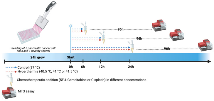
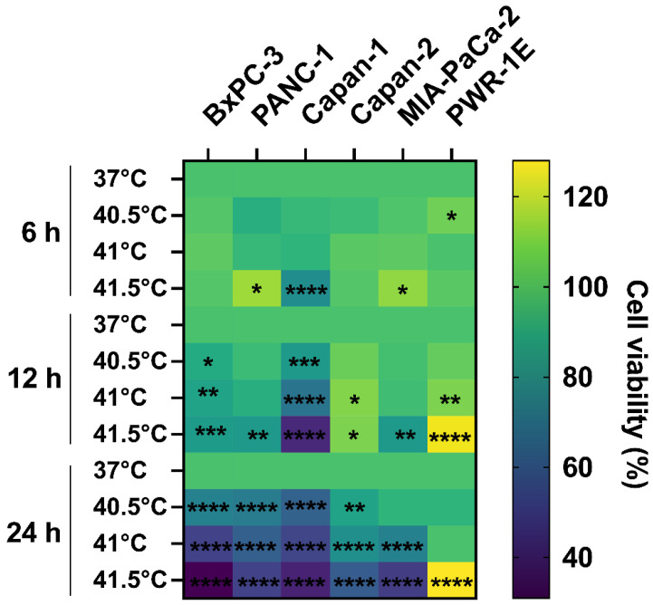
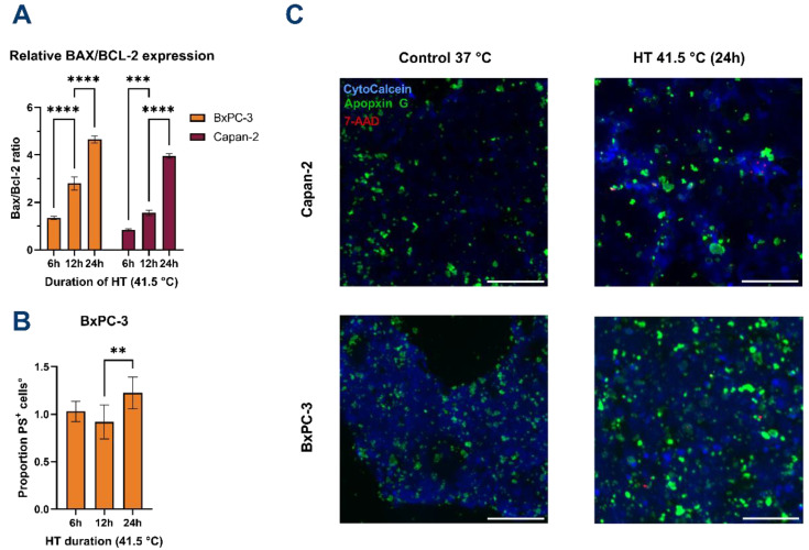
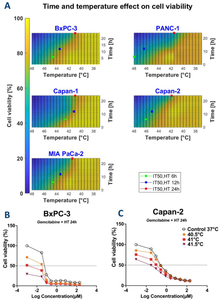
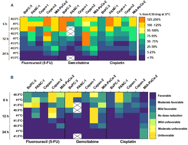
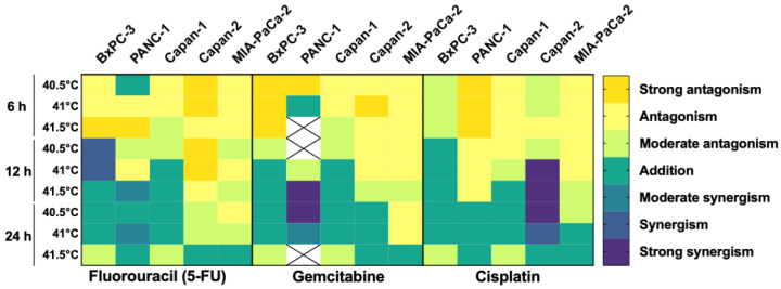
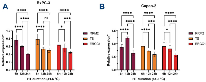
Chemotherapy (CT) is the standard care for advanced pancreatic ductal adenocarcinoma (PDAC); however, with limited efficacy. Hyperthermia (HT) treatment has been suggested as a sensitizer to improve outcomes. However, the direct effect of the HT and CT combination is not fully understood. Therefore, we aim to assess the direct cytotoxic effect of HT in PDAC cells as monotherapy or in combination with chemotherapeutics. Different temperatures (37-, 40.5-, 41-, and 41.5 °C) and durations (6-, 12-, and 24 h) were tested in PDAC cell lines (BxPC-3, Capan-1, Capan-2, PANC-1, and MIA-PaCa-2). Different concentrations of gemcitabine, 5-fluorouracil, and cisplatin were also tested in these conditions. The impact on cell metabolic activity was determined by an MTS assay. Enhancement of chemosensitivity was assessed by a reduction in half-maximal inhibitory concentration (IC50). HT and chemotherapeutics interactions were classified as antagonistic, additive, or synergistic using the combination index. HT inhibited cell proliferation in a cell type, temperature, and duration-dependent manner. The induction of apoptosis was seen after 6 h of HT treatment, eventually followed by secondary necrosis. The HT and CT combination led to an IC50 reduction of the tested CT. At 12 h of HT, this effect was between 25 to 90% and reached a 95% reduction at 24 h. The additive or synergistic effect was demonstrated in all cell lines and chemotherapeutics, although, again, this depended on cell type, duration, and temperature. HT is cytotoxic and enhances the therapeutic effectiveness of gemcitabine, 5-fluorouracil, and cisplatin on PDAC cells. This result was further confirmed by the decrease in the expression of RRM2, TS, and ERCC1 in BxPC-3 and Capan-2 cells. These observations warrant further study in specific subsets of PDAC patients to improve their clinical outcomes.

Keywords: 5-fluorouracil; anticancer therapy; cell proliferation; cisplatin; gemcitabine; thermal therapy.


Conflict of interest statement

The authors declare no conflict of interest.

Figures

Figure 1
Figure 1
Experimental design. Pancreatic tumor cell lines (BxPC-3, Capan-1, Capan-2, PANC-1, and MIA-PaCa-2) were seeded and expanded for 24 h. Gemcitabine, 5-fluorouracil, and cisplatin were added immediately after hyperthermia treatment using different temperatures (40.5-, 41-, and 41.5 °C) and durations (6-, 12-, and 24 h). Controls were kept at 37 °C. After 96 h, the influence of chemotherapy and temperature on cell survival was determined by MTS assay. Created with BioRender.com.
Figure 2
Figure 2
Effect of hyperthermia on pancreatic cancer cells. Heatmap showing the effect of hyperthermia on different cell lines for different durations (6, 12, and 24 h) and different temperatures (37 °C = control, 40.5-, 41-, and 41.5 °C). The color legend shows the percentage of relative cell survival compared to controls. There is a general trend of increased cell death with increased temperature and time of exposure. The healthy control cells are not killed by hyperthermia treatment. Results were expressed as relative cell viability as compared to 37 °C. Statistical significance * p < 0.05; ** p < 0.001; *** p < 0.0001; **** p < 0.00001.
Figure 3
Figure 3
Hyperthermia induces apoptosis in PDAC cells. (A) BAX/BCL-2 expression ratio significantly increases over HT duration for both cell lines. Values were normalized for GAPDH expression and calculated relative to controls at 37 °C. (B) Expression of phosphatidylserine (PS) on the outer membrane is significantly increased after 24 h of HT only for BxPC-3. Data are normalized to controls at 37 °C. Data for Capan-2 are shown in Supplementary Figure S1. No significant differences in PS positivity were observed for Capan-2. (C) Representative fluorescence microscopy imaging of BxPC-3 and Capan-2 cells at 37 °C (control) and after 24 h at 41.5 °C. Viable cells are stained blue, apoptotic cells are stained green (PS), and necrotic nuclei are stained red. Images were acquired at 4× magnification. Scale bars represent 200 µm. All error bars represent 95% CI. Statistical significance ** p < 0.001; *** p < 0.0001; **** p < 0.00001.
Figure 4
Figure 4
Combination of time and temperature effects on cell proliferation as illustrated by heatmaps for the calculation of IT50,HT and IC50,drug for thermal therapy (A) Heatmaps showing the relative cell viability in function of time and temperature were used to calculate IT50,HT for thermal therapy. IT50,HT values are shown for each of the cell lines at 6-, 12-, and 24 h. Heatmaps show that treatment duration increases the cytotoxic effect of thermal therapy observed by a lower IT50,HT (i.e., lower temperatures are needed to decrease cell viability by half of its maximal effect). (B,C): Dose curve response of BxPC-3 and Capan-2 to gemcitabine for 24 h.
Figure 5
Figure 5
Hyperthermia enhances the cytotoxic effect of chemotherapeutic agents. Heatmap displaying the IC50,ratio, drug which represent the combined effect of hyperthermia and chemotherapy compared to single therapy (drug administered at 37 °C). Data are expressed as a percentage of IC50,drug at 37 °C experimental conditions. X indicates data not available (A). Dose reduction index values are plotted in (B) to further confirm the results expressed as relative IC50,ratio, drug. Unfavorable: DRI < 0.8; Moderate unfavorable: 0.8 < DRI > 0.85; Mild unfavorable: 0.85 < DRI > 0.9; No dose reduction: 0.9 < DRI > 1.1; Mild Favorable: 1.1 < DRI > 1.3; Moderate favorable 1.3 < DRI > 1.8; Favorable DRI > 1.8. X indicates data not available.
Figure 6
Figure 6
Hyperthermia and chemotherapeutics have additive/synergistic anticancer effects. Heatmap showing the combination index of hyperthermia and chemotherapy using different pancreatic cell lines and different temperatures and durations of hyperthermia. The combined effect between the two therapies is expressed as antagonistic, additive, or synergistic. Strong synergism: CI < 0.8; Synergism: 0.8 < CI > 0.85; Moderate synergism: 0.85 < CI > 0.9; Addition: 0.9 < CI > 1.1; Moderate antagonism: 1.1< CI >1.3; Antagonism 1.3< CI >1.8; Strong Antagonism CI > 1.9. X indicates data not available.
Figure 7
Figure 7
Hyperthermia leads to downregulation of RRM2, TS, and ERCC1. Relative expression of RRM1, TS, and ERCC1 in (A) BxPC-3 cells and (B) Capan-2 cells. Expression is normalized to GAPDH and calculated relative to cells incubated at 37 °C. Statistical significance ns = not significant; * p < 0.05; *** p < 0.0001; **** p < 0.00001.

References

    1. Cai J., Chen H., Lu M., Zhang Y., Lu B., You L., Zhang T., Dai M., Zhao Y. Advances in the epidemiology of pancreatic cancer: Trends, risk factors, screening, and prognosis. Cancer Lett. 2021;520:1–11. doi: 10.1016/j.canlet.2021.06.027. - DOI - PubMed
    1. Tempero M.A. NCCN Guidelines Updates: Pancreatic Cancer. J. Natl. Compr. Cancer Netw. 2019;17:603–605. doi: 10.6004/jnccn.2019.5007. - DOI - PubMed
    1. Milella M., Bassi C., Boggi U., Brunetti O., Cavaliere A., Crippa S., De Vita F., Falconi M., Frassineti G.L., Giommoni E., et al. Evolving pancreatic cancer treatment: From diagnosis to healthcare management. Crit. Rev. Oncol. 2021;169:103571. doi: 10.1016/j.critrevonc.2021.103571. - DOI - PubMed
    1. Beatty G.L., Werba G., Lyssiotis C.A., Simeone D.M. The biological underpinnings of therapeutic resistance in pancreatic cancer. Genes Dev. 2021;35:940–962. doi: 10.1101/gad.348523.121. - DOI - PMC - PubMed
    1. Quintiliani M. Review the Oxygen Effect in Radiation Inactivation of DNA and Enzymest. Int. J. Radiat. Biol. Relat. Stud. Phys. Chem. Med. 1986;50:573–594. doi: 10.1080/09553008614550981. - DOI - PubMed

MeSH terms