. 2022 May 23;12(5):734.
doi: 10.3390/biom12050734.

Impairment of the Retinal Endothelial Cell Barrier Induced by Long-Term Treatment with VEGF-A165 No Longer Depends on the Growth Factor's Presence

Affiliations

Impairment of the Retinal Endothelial Cell Barrier Induced by Long-Term Treatment with VEGF-A165 No Longer Depends on the Growth Factor's Presence

Heidrun L Deissler et al. Biomolecules. .

Abstract

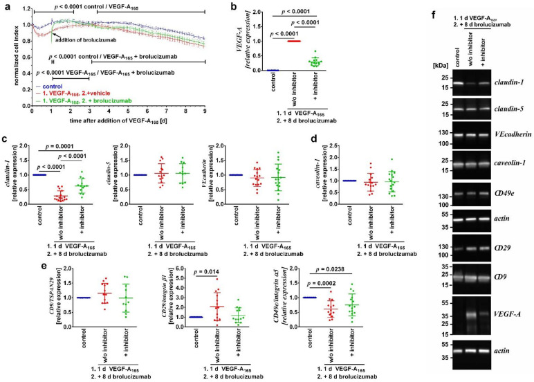
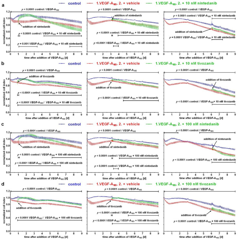
As responses of immortalized endothelial cells of the bovine retina (iBREC) to VEGF-A165 depend on exposure time to the growth factor, we investigated changes evident after long-term treatment for nine days. The cell index of iBREC cultivated on gold electrodes-determined as a measure of permeability-was persistently reduced by exposure to the growth factor. Late after addition of VEGF-A165 protein levels of claudin-1 and CD49e were significantly lower, those of CD29 significantly higher, and the plasmalemma vesicle associated protein was no longer detected. Nuclear levels of β-catenin were only elevated on day two. Extracellular levels of VEGF-A-measured by ELISA-were very low. Similar to the binding of the growth factor by brolucizumab, inhibition of VEGFR2 by tyrosine kinase inhibitors tivozanib or nintedanib led to complete, although transient, recovery of the low cell index when added early, though was inefficient when added three or six days later. Additional inhibition of other receptor tyrosine kinases by nintedanib was similarly unsuccessful, but additional blocking of c-kit by tivozanib led to sustained recovery of the low cell index, an effect observed only when the inhibitor was added early. From these data, we conclude that several days after the addition of VEGF-A165 to iBREC, barrier dysfunction is mainly sustained by increased paracellular flow and impaired adhesion. Even more important, these changes are most likely no longer VEGF-A-controlled.

Keywords: VEGF-A; cell adhesion; claudin-1; nintedanib; paracellular flow; plasmalemma vesicle associated protein; retinal endothelial cells; tight junction; tivozanib; transcellular flow.


Conflict of interest statement

H.L.D. has no conflicts of interest to declare. M.R. is consultant for Allergan, Alimera, Bayer and Novartis, received speaker honoraria from Allergan, Alimera, Bayer, Novartis, and Zeiss. A.W. received financial support from Alimera and Bayer and speaker honoraria from Alimera, Allergan, Bayer, Novartis, and Oertli.

Figures

Figure 1
Figure 1
Prolonged treatment with VEGF-A165 impaired the barrier formed by iBREC. Confluent monolayers of iBREC were exposed to VEGF-A165 for nine days and (a) the cell index was measured continuously or (bf) cells were harvested. Alternatively, iBREC were treated with the growth factor for one day, before the VEGF-A antagonist brolucizumab was added for an additional eight days. (a) VEGF-A165 induced a persistent decrease in the cell index, which was quickly reverted by brolucizumab, but this effect was only stable for 1.5 days. Cell index values obtained from more than five wells per condition were normalized to those measured just before the addition of VEGF-A165 and are shown as means ± standard deviations. Data were analyzed as described in “Materials and Methods”. (b) Less VEGF-A was taken up by iBREC in the presence of brolucizumab. (c) Levels of TJ-protein claudin-1 were low after exposure of the cells to the growth factor in the absence or presence of its antagonist. (d) None of the effectors changed expression levels of caveolin-1. (e) VEGF-A165 induced higher levels of CD29/integrin α1 but lower levels of CD49e/integrin α5 not changed by brolucizumab. Signals obtained by Western blot analyses were normalized and statistical analyses performed as described in “Materials and Methods”. Pooled results of multiple Western blot experiments are shown as scatter plots also depicting means ± standard deviations where one dot represents the single signal obtained from one experiment. (f) Representative images of Western blot analyses; the original images are shown in Figure S1.
Figure 2
Figure 2
Nuclear expression levels of β-catenin changed over time of exposure to VEGF-A165. Confluent iBREC were exposed to VEGF-A165 and cells were harvested one to six days later for preparation of (a) cellular and (b) nuclear extracts and subsequent Western blot analyses of which representative images are shown in (c). Levels of (a) cellular β-catenin remained unchanged by VEGF-A165 but (b) those of nuclear β-catenin were significantly elevated only on day 2. Signals obtained by Western blot analyses were normalized and analyzed as described above. Please refer to Figure S2 for original images.
Figure 3
Figure 3
Plasma membrane localization of β-catenin was impaired by extended exposure of iBREC to VEGF-A165. Confluent cells were exposed to VEGF-A165 and cells were fixated at indicated time points. Subcellular localization of β-catenin was visualized by immunofluorescence stainings with specific antibodies (red); nuclei were stained with DAPI (blue). In unchallenged cells, most of β-catenin was present at the plasma membrane, but exposure to VEGF-A165 for one to three days resulted in its de-localization from the plasma membrane, less evident after extended exposure for six days. Scale bar 50 µm.
Figure 4
Figure 4
Inhibition of only VEGFR2 was not sufficient to normalize late VEGF-A165-induced barrier dysfunction. After exposure of confluent monolayers of iBREC cultivated on gold electrodes to VEGF-A165, (a,b) 10 nM or (c,d) 100 nM, nintedanib or tivozanib were added at indicated time points, and the cell index was measured continuously. Cell index values obtained from more than five wells per condition were normalized to those measured just before the addition of VEGF-A165 and are shown as means ± standard deviations. (a,b) Only when added during the early phase of VEGF-A-exposure were 10 nM nintedanib or tivozanib able to revert the induced changes, although these effects were not long-lasting. (c,d) When added during the early phase of VEGF-A-exposure, tivozanib, but not nintedanib, completely and sustainably normalized induced changes. Both inhibitors had little or no effect when added later.

References

    1. Aiello L.P., Avery R.L., Arrigg P.G., Keyt B.A., Jampel H.D., Shah S.T., Pasquale L.R., Thieme H., Iwamoto M.A., Park J.E., et al. Vascular endothelial growth factor in ocular fluid of patients with diabetic retinopathy and other retinal disorders. N. Engl. J. Med. 1994;331:1480–1487. doi: 10.1056/NEJM199412013312203. - DOI - PubMed
    1. Campochiaro P.A. Retinal and choroidal neovascularization. J. Cell Physiol. 2000;184:301–310. doi: 10.1002/1097-4652(200009)184:3<301::AID-JCP3>3.0.CO;2-H. - DOI - PubMed
    1. Campochiaro P.A. Ocular neovascularization. J. Mol. Med. 2013;91:311–321. doi: 10.1007/s00109-013-0993-5. - DOI - PMC - PubMed
    1. Uemura A., Fruttiger M., D’Amore P.A., De Falco S., Joussen A.M., Sennlaub F., Brunck L.R., Johnson K.T., Lambrou G.N., Rittenhouse K.D., et al. VEGFR1 signaling in retinal angiogenesis and microinflammation. Prog. Retin. Eye Res. 2021;84:100954. doi: 10.1016/j.preteyeres.2021.100954. - DOI - PMC - PubMed
    1. Antonetti D.A., Barber A.J., Hollinger L.A., Wolpert E.B., Gardner T.W. Vascular endothelial growth factor induces rapid phosphorylation of tight junction proteins occludin and zonula occludens 1. J. Biol. Chem. 1999;274:23463–23467. doi: 10.1074/jbc.274.33.23463. - DOI - PubMed

MeSH terms

Substances