Review
. 2022 Apr 22;10(5):974.
doi: 10.3390/biomedicines10050974.

Controlling Cancer Cell Death Types to Optimize Anti-Tumor Immunity

Affiliations
Review

Controlling Cancer Cell Death Types to Optimize Anti-Tumor Immunity

Marie Oliver Metzig et al. Biomedicines. .

Abstract

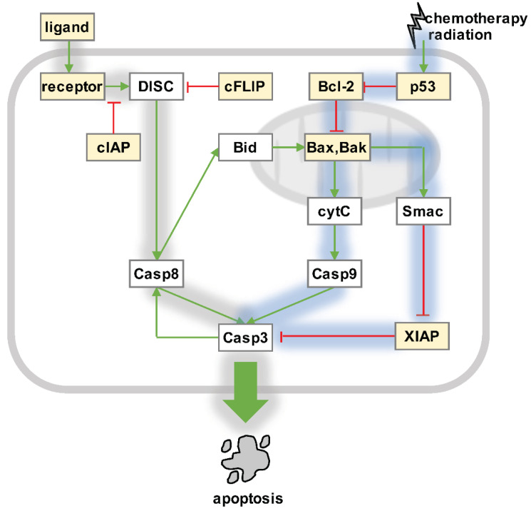
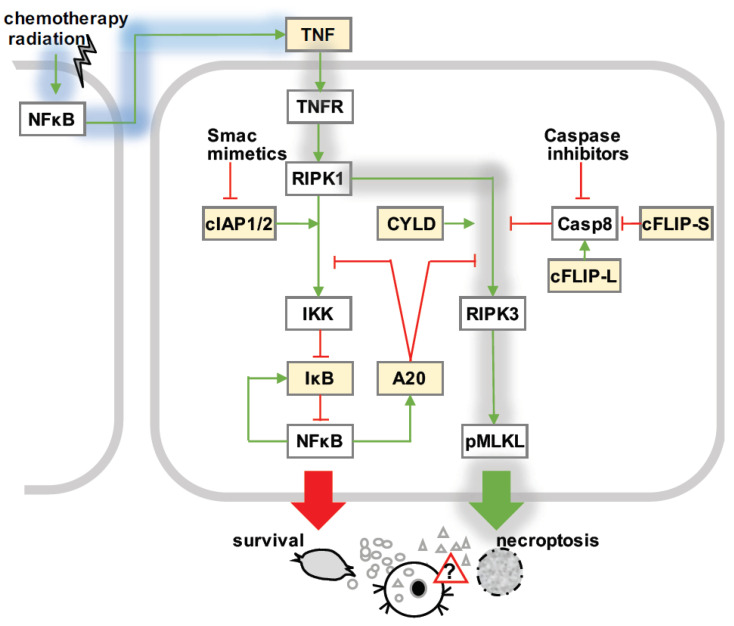
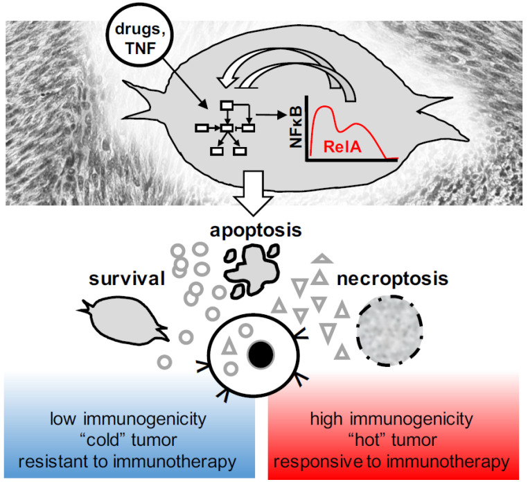
Over several decades, cell biology research has characterized distinct forms of regulated cell death, identified master regulators such as nuclear factor kappa B (NFκB), and contributed to translating these findings in order to improve anti-cancer therapies. In the era of immunotherapy, however, the field warrants a new appraisal-the targeted induction of immunogenic cell death may offer personalized strategies to optimize anti-tumor immunity. Once again, the spotlight is on NFκB, which is not only a master regulator of cancer cell death, survival, and inflammation, but also of adaptive anti-tumor immune responses that are triggered by dying tumor cells.

Keywords: NFκB dynamics; anti-tumor immunity; fate decisions; immunogenic cell death; immunotherapy.


Conflict of interest statement

The authors declare no conflict of interest.

Figures

Figure 1
Figure 1
Apoptosis signaling. Intrinsic pathway (blue shading): DNA damage is sensed by tumor suppressor p53. P53 controls the Bcl-2 protein family, which includes pro-apoptotic (e.g., Bid and Bax) and pro-survival (e.g., Bcl-2) factors that tightly regulate mitochondrial outer membrane permeabilization (MOMP). Activated Bax and Bak form pores in the outer mitochondrial membranes, which allow Smac/Diablo and cytochrome c to translocate from the intermembrane space into the cytosol. Cytochrome c binds Apaf-1 and caspase 9, which form the so-called “apoptosome” to activate effector caspases 3, 6, and 7. Smac/Diablo inhibits XIAP, which releases the block on the proteolytic activity of effector caspases. Extrinsic pathway (gray shading): Pro-apoptotic ligands such as TNF, FasL, or TRAIL engage death receptors on tumor cells, leading to the formation of death-inducing signaling complex (DISC) by recruiting FADD and the pro-forms of initiator caspases 8 and 10. Active initiator caspases may then activate effector caspases 3, 6, and 7, but this is limited in most cancer cells because of inhibitory factors such as cIAP, cFLIP, and XIAP. Caspase 8 may engage the intrinsic pathway via Bid truncation and MOMP. Released effector caspases may create a positive feedback and activate more caspase 8 in some cells. NFκB-responsive regulators (yellow boxes): The molecular network of apoptosis regulation includes NFκB target genes on several levels. Changes in NFκB activities therefore determine the abundances of pro-survival and pro-apoptosis regulators, and affect tumor cell fate decisions in response to death-ligands, chemotherapy, and radiation.
Figure 2
Figure 2
Necroptosis signaling. TNF-mediated necroptosis signaling (gray shading): Upon TNF stimulation, rapid recruitment of RIPK1 to TNFR1 (complex I) leads to canonical activation of NFκB via the inhibitor kB kinase (IKK), which targets the inhibitor of κB (IκB) proteins for phosphorylation and subsequent degradation. After a delay period, RIPK1 dissociates from plasma-membrane-bound complex I to bind to RIPK3 (complex IIb or necrosome). Activated RIPK3 recruits and phosphorylates MLKL (pMLKL), which executes necroptotic cell death. cIAP1 and cIAP2 stabilize complex I, while CYLD facilitates the formation of the necrosome. A20 may hamper the activation of IKK via complex I, or inhibit the activation of RIPK3 in the necrosome. The ratio of long and short cFLIP isoforms relative to caspase 8 control the activity of caspase 8: pro-caspase-8-cFLIP-L, but not –cFLIP-S heterodimers destabilize the necrosome. Necroptosis is facilitated by caspase inhibitors, and Smac mimetics, which target cIAP1 and cIAP2 for degradation. NFκB-responsive regulators (yellow boxes): Basal and inducible NFκB is not only known to control the gene expression programs regulating necroptosis decisions (yellow), but may also affect the immunogenicity of dying cells. Cancer therapies trigger NFκB via DNA damage and initiate auto- or para-crine TNF signaling, which mediates tumor cell survival or death.
Figure 3
Figure 3
Dynamic apoptosis and necroptosis decisions and their consequences for tumor immunity. NFκB is a dynamic regulator of the molecular network that governs over cell death and survival, as well as the immunological consequences of dying tumor cells. Immunogenic cell death induced by anti-cancer therapies may convert a subset of “cold” tumors into “hot” tumors, and thus help to overcome resistance to immunotherapy.

Similar articles

References

    1. Hanahan D., Weinberg R.A. Hallmarks of cancer: The next generation. Cell. 2011;144:646–674. doi: 10.1016/j.cell.2011.02.013. - DOI - PubMed
    1. Ashkenazi A. Directing cancer cells to self-destruct with pro-apoptotic receptor agonists. Nat. Rev. Drug Discov. 2008;7:1001–1012. doi: 10.1038/nrd2637. - DOI - PubMed
    1. Galluzzi L., Vitale I., Aaronson S.A., Abrams J.M., Adam D., Agostinis P., Alnemri E.S., Altucci L., Amelio I., Andrews D.W., et al. Molecular mechanisms of cell death: Recommendations of the Nomenclature Committee on Cell Death 2018. Cell Death Differ. 2018;25:486–541. doi: 10.1038/s41418-017-0012-4. - DOI - PMC - PubMed
    1. von Karstedt S., Montinaro A., Walczak H. Exploring the TRAILs less travelled: TRAIL in cancer biology and therapy. Nat. Rev. Cancer. 2017;17:352–366. doi: 10.1038/nrc.2017.28. - DOI - PubMed
    1. Fulda S. Promises and Challenges of Smac Mimetics as Cancer Therapeutics. Clin. Cancer Res. An Off. J. Am. Assoc. Cancer Res. 2015;21:5030–5036. doi: 10.1158/1078-0432.CCR-15-0365. - DOI - PubMed