. 2022 May 3;10(5):1056.
doi: 10.3390/biomedicines10051056.

Hypoxia Induces DPSC Differentiation versus a Neurogenic Phenotype by the Paracrine Mechanism

Affiliations

Hypoxia Induces DPSC Differentiation versus a Neurogenic Phenotype by the Paracrine Mechanism

Simona Delle Monache et al. Biomedicines. .

Abstract

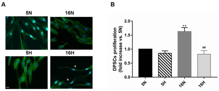
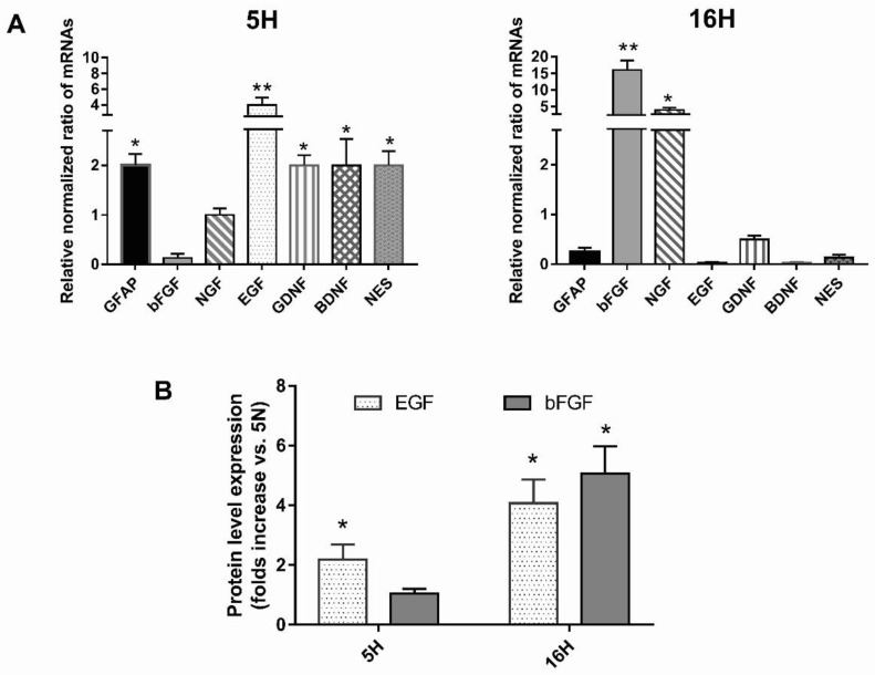
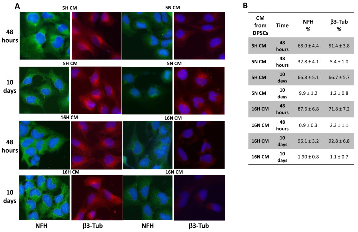
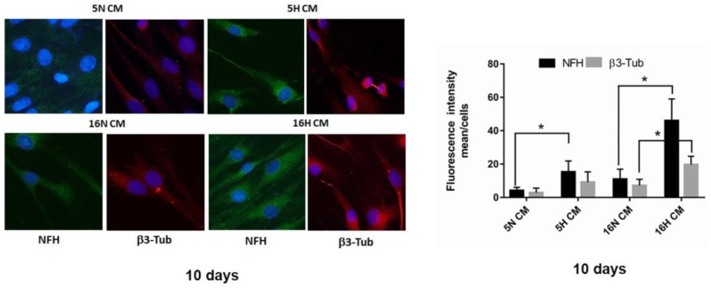
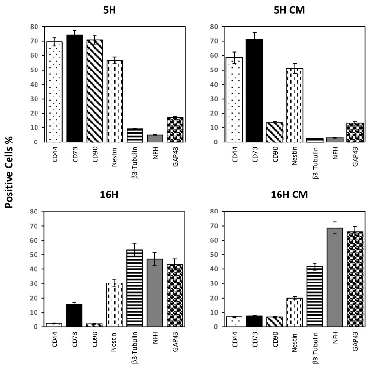
As previously described by several authors, dental pulp stem cells (DPSCs), when adequately stimulated, may acquire a neuronal-like phenotype acting as a favorable source of stem cells in the generation of nerves. Besides, it is known that hypoxia conditioning is capable of stimulating cell differentiation as well as survival and self-renewal, and that multiple growth factors, including Epidermal Growth factor (EGF) and basic fibroblast growth factor (bFGF), are often involved in the induction of the neuronal differentiation of progenitor cells. In this work, we investigated the role of hypoxia in the commitment of DPSCs into a neuronal phenotype. These cells were conditioned with hypoxia (O2 1%) for 5 and 16 days; subsequently, we analyzed the proliferation rate and morphology, and tested the cells for neural and stem markers. Moreover, we verified the possible autocrine/paracrine role of DPSCs in the induction of neural differentiation by comparing the secretome profile of the hypoxic and normoxic conditioned media (CM). Our results showed that the hypoxia-mediated DPSC differentiation was time dependent. Moreover, conditioned media (CM derived from DPSCs stimulated by hypoxia were able, in turn, to induce the neural differentiation of SH-SY5Y neuroblastoma cells and undifferentiated DPSCs. In conclusion, under the herein-mentioned conditions, hypoxia seems to favor the differentiation of DPSCs into neuron-like cells. In this way, we confirm the potential clinical utility of differentiated neuronal DPSCs, and we also suggest the even greater potential of CM-derived-hypoxic DPSCs that could more readily be used in regenerative therapies.

Keywords: DPSCs; dental pulp; hypoxia; neuronal differentiation; stem cells.


Conflict of interest statement

The authors declare no conflict of interest.

Figures

Figure 1
Figure 1
Effects of hypoxia on DPSC morphology and proliferation. (A) Representative images of phalloidin-stained DPSCs exposed or not to hypoxia for 5 and 16 days, as described in the Materials and Methods section (Table 1). White arrows indicate cell morphology changes. Scale bar 20 µm. (B) Histograms show that hypoxia induced a significant decrease in proliferation rate exclusively after 16 days of exposure in comparison to normoxia. Histograms indicate the means ± SE of three different cultures, each of which was tested in triplicate. Statistical analysis was performed by one-way ANOVA followed by Tukey’s test. ∗∗ p < 0.01 vs. 5N, and ## p < 0.01 vs. 16N.
Figure 2
Figure 2
Effects of hypoxia on phenotypic expression profile of DPSCs. (A) shows that mesenchymal stem cells markers CD44, CD90, CD105, STRO1 and CD73 decreased in cells exposed to hypoxia at 5 and 16 days of exposure. Conversely, neuronal markers increased after hypoxia exposure. Phenotypic expression of 16H was very similar to (B) DPSCs treated with EGF and bFGF factors for 14 days, a culture condition that determines the neuronal differentiation of DPSCs.
Figure 3
Figure 3
Effects of hypoxia on the mRNA profile expression of DPSCs. (A) Real-time RT-qPCR analysis of the levels of expression of early and late markers of neural differentiation (nestin, GFAP, EGF, NGF, bFGF BDNF, GDNF) after 5 days and after 16 days of hypoxic exposure. (B) Secreted growth factors bFGF and EGF were detected in the conditioned media of DPSCs exposed or not exposed to hypoxia for 5 days and 16 days by human EGF and FGF basic ELISA Kits. Histograms indicate the means ± SE of three different cultures, each of which was tested in triplicate. Statistical analysis was performed by Student’s t-test; * p < 0.05; ** p < 0.01 vs. 5N.
Figure 4
Figure 4
Immunofluorescence and flow cytometry analysis of SH-SY5Y. (A) SH-SY5Y exposed to DPSC-CM (5H, 16H) were stained for neuronal markers NFH and β3-Tubulin that were highly expressed in all groups of SH-SY5Y treated with DPSCs’ hypoxic CM (5H, 16H) in comparison with groups of cells treated with DPSCs’ normoxic CM (5N, 16N). (B) Flow cytometry analysis of NFH and β3-Tubulin expression in SH-SY5Y-treated groups or untreated cells. Scale bar 20 µm.
Figure 5
Figure 5
Immunofluorescence analysis of DPSCs. (left). Immunofluorescence analysis of NFH and β3-Tubulin in DPSCs treated with DPSC-CM (5H CM, 16H CM) or DPSC-CM (5N CM, 16N CM) groups. Scale bar 20 µm. (right). Analysis of fluorescence intensity NFH and β3-Tubulin in DPSCs treated with DPSC-CM (5H CM, 16H CM) or DPSC-CM (5N CM, 16N CM) groups. Data are represented as mean ± SE. Statistical analysis was performed by Student’s t-test; * p < 0.05; 5H and 16H vs. 5N and 16N.
Figure 6
Figure 6
Effects of hypoxia on the phenotypic expression profiles of DPSCs treated with CM enriched by hypoxia. Flow cytometry analysis of DPSC-treated groups; 5H CM and 16H CM were compared with the 5H and 16H groups to determine the profile of expression of several neuronal and stem markers. Histograms indicate the means ± SE of three different experiments.

References

    1. Al Madhoun A., Sindhu S., Haddad D., Atari M., Ahmad R., Al-Mulla F. Dental Pulp Stem Cells Derived From Adult Human Third Molar Tooth: A Brief Review. Front. Cell Dev. Biol. 2021;9:717624. doi: 10.3389/fcell.2021.717624. - DOI - PMC - PubMed
    1. Diomede F., Fonticoli L., Marconi G.D., Della Rocca Y., Rajan T.S., Trubiani O., Murmura G., Pizzicannella J. Decellularized Dental Pulp, Extracellular Vesicles, and 5-Azacytidine: A New Tool for Endodontic Regeneration. Biomedicines. 2022;10:403. doi: 10.3390/biomedicines10020403. - DOI - PMC - PubMed
    1. Tatullo M., Marrelli M., Shakesheff K.M., White L.J. Dental pulp stem cells: Function, isolation and applications in regenerative medicine. J. Tissue Eng. Regen. Med. 2015;9:1205–1216. doi: 10.1002/term.1899. - DOI - PubMed
    1. Anitua E., Troya M., Zalduendo M. Progress in the use of dental pulp stem cells in regenerative medicine. Cytotherapy. 2018;20:479–498. doi: 10.1016/j.jcyt.2017.12.011. - DOI - PubMed
    1. Brown C., McKee C., Bakshi S., Walker K., Hakman E., Halassy S., Svinarich D., Dodds R., Govind C.K., Chaudhry G.R. Mesenchymal stem cells: Cell therapy and regeneration potential. J. Tissue Eng. Regen. Med. 2019;13:1738–1755. doi: 10.1002/term.2914. - DOI - PubMed