Review
. 2022 May 4;10(5):1065.
doi: 10.3390/biomedicines10051065.

(Dis)similarities between the Decidual and Tumor Microenvironment

Affiliations
Review

(Dis)similarities between the Decidual and Tumor Microenvironment

Jelena Krstic et al. Biomedicines. .

Abstract

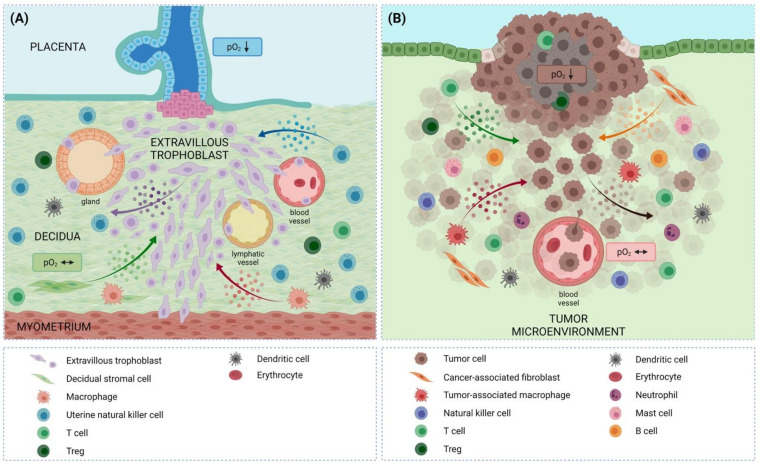
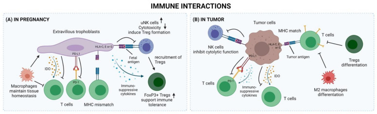
Placenta-specific trophoblast and tumor cells exhibit many common characteristics. Trophoblast cells invade maternal tissues while being tolerated by the maternal immune system. Similarly, tumor cells can invade surrounding tissues and escape the immune system. Importantly, both trophoblast and tumor cells are supported by an abetting microenvironment, which influences invasion, angiogenesis, and immune tolerance/evasion, among others. However, in contrast to tumor cells, the metabolic, proliferative, migrative, and invasive states of trophoblast cells are under tight regulatory control. In this review, we provide an overview of similarities and dissimilarities in regulatory processes that drive trophoblast and tumor cell fate, particularly focusing on the role of the abetting microenvironments.

Keywords: decidual microenvironment; immune cells; invasion; placenta; proliferation; trophoblast; tumor cell; tumor microenvironment.


Conflict of interest statement

The authors declare no conflict of interest.

Figures

Figure 1
Figure 1
Comparison between the decidual and the tumor microenvironment. Invasive trophoblasts and tumor cells are both sustained by an abetting microenvironment. Intense crosstalk between the extravillous trophoblast/tumor cells and the cells of their microenvironment, in particular immune cells (e.g., via the secretion of molecules depicted as colored dots for certain cell types), is essential for establishing and maintaining immune tolerance/suppression towards extravillous trophoblast/tumor cells. Furthermore, cells of the microenvironment, including immune cells, are involved in regulating invasion and angiogenesis in both settings. (A) On the left, extravillous trophoblasts invade the uterine tissues of the mother, reaching the inner third of the myometrium. The extravillous trophoblasts invade the decidual stroma, vessels, and glands—all potential sources of nutrients. During the first trimester of pregnancy, the oxygen concentration within the villous part of the placenta has been found to be below 20 mmHg. Trophoblasts proliferate in this low-oxygen environment. From this physiologically low oxygen level (normoxia for the placenta at this stage of pregnancy), extravillous trophoblasts invade normally oxygenated uterine tissues and thus follow an oxygen gradient towards higher levels. (B) On the right, tumor cells can similarly invade surrounding tissues and again follow an oxygen gradient towards higher oxygen levels. Similar to extravillous trophoblasts, tumor cells proliferate in the peripheral zones around sites of low oxygen (real hypoxia), while subsequent migration and invasion take place towards higher oxygen levels. Created with BioRender.com (accessed on 23 April 2022).
Figure 2
Figure 2
Comparison of the immune interactions found in (A) the decidual microenvironment and (B) the tumor microenvironment. In both, similar immune cells (NK cells, T cells, Tregs, and macrophages) are found. Both may also use similar mechanisms of immune evasion to suppress the immune responses of the host, mediated by the secretion of immunosuppressive cytokines, metabolites (e.g., IDO), co-inhibitory signals, non-classical MHC class I molecules and/or by the recruitment of Tregs and/or NK cells. Secreted factors are shown as dots. Created with BioRender.com (accessed on 23 April 2022).

References

    1. Benirschke K., Burton G.J., Baergen R.N. Pathology of the Human Placenta. 6th ed. Springer; Berlin/Heidelberg, Germany: 2012.
    1. Maltepe E., Bakardjiev A.I., Fisher S.J. The placenta: Transcriptional, epigenetic, and physiological integration during development. J. Clin. Investig. 2010;120:1016–1025. doi: 10.1172/JCI41211. - DOI - PMC - PubMed
    1. Yang F., Zheng Q., Jin L. Dynamic Function and Composition Changes of Immune Cells During Normal and Pathological Pregnancy at the Maternal-Fetal Interface. Front. Immunol. 2019;10:2317. doi: 10.3389/fimmu.2019.02317. - DOI - PMC - PubMed
    1. Sharma S., Godbole G., Modi D. Decidual Control of Trophoblast Invasion. Am. J. Reprod. Immunol. 2016;75:341–350. doi: 10.1111/aji.12466. - DOI - PubMed
    1. Bezemer R.E., Schoots M.H., Timmer A., Scherjon S.A., Erwich J.J.H.M., van Goor H., Gordijn S.J., Prins J.R. Altered Levels of Decidual Immune Cell Subsets in Fetal Growth Restriction, Stillbirth, and Placental Pathology. Front. Immunol. 2020;11:1898. doi: 10.3389/fimmu.2020.01898. - DOI - PMC - PubMed

LinkOut - more resources